Over 1.71 billion people globally suffer from musculoskeletal conditions. Such disorders affect the muscles, bones, and joints, significantly reducing mobility and productivity. According to the World Health Organisation, disabilities related to musculoskeletal conditions have been increasing and are projected to continue to rise in the next decades.
Early intervention is a proactive measure to spot early signs of an injury or condition and help you heal faster. In most cases, standard imaging like x-rays and regular MRIs don’t show enough detail to resolve the problem.
That’s when you might require an arthrogram. Vista Health offers MR arthrogram scans in London and 2 other clinics across England, that help health providers detect abnormalities and plan appropriate treatment. In this guide, we’ll discuss what happens before, during and after an arthrogram procedure.
What is an arthrogram?
An arthrogram is a type of imaging test used to inspect the inside of a joint such as a shoulder, wrist, knee or ankle to properly diagnose an injury or a disorder. The test consists of two parts: injection and MRI.
During an MR arthrogram procedure, the tendon, cartilage, or ligament is examined for signs of disease, tears, or degeneration. It’s also conducted to identify growths or synovial cysts in the joint and further evaluate issues detected in a prior MRI.
Preparation
No specific preparation is required for an MR arthrogram. That said, it pays to wear comfortable, loose-fitting clothing without metal fasteners, such as buttons, snaps and zippers.
During the scan
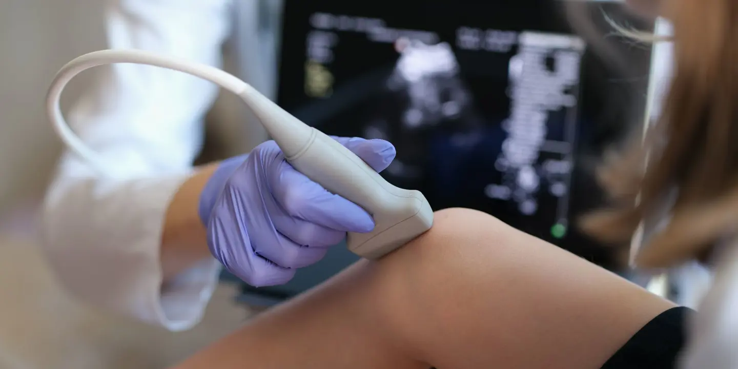
First, you’ll be asked to take off any jewellery and clothing around the joint and lay on a table in the exam room. Using an antiseptic solution, the skin over the joint will be carefully sanitised. Then a local anesthetic may be injected to numb the area where the dye will be administered.
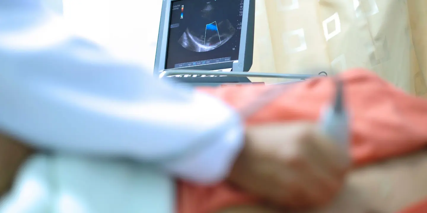
Dye or contrast medium will be injected into the joint. This helps produce clearer images of soft tissue structures, allowing your doctor to come up with a more accurate diagnosis. An x-ray or ultrasound scan will be used to ensure the dye is injected in the right area.
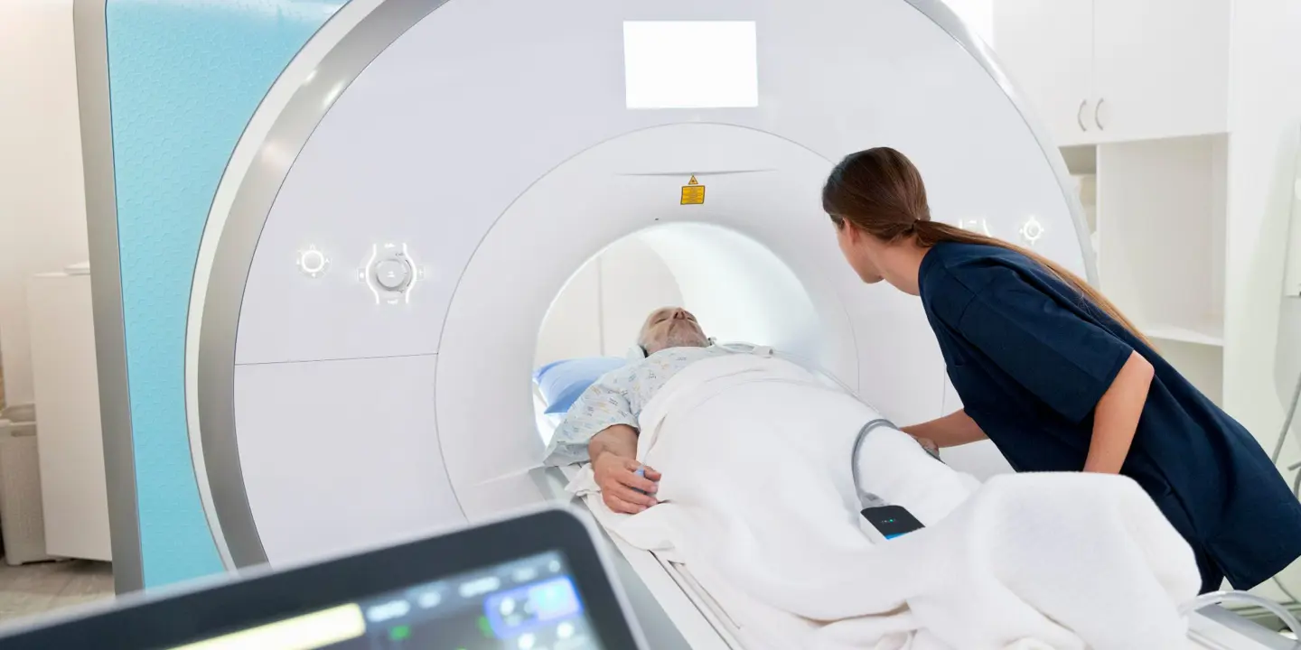
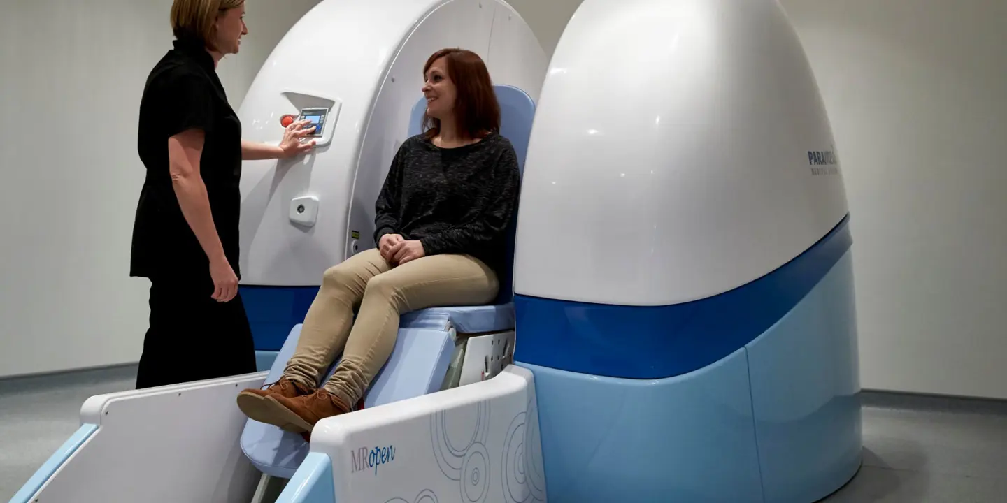
After the injections, you will proceed to the MRI suite where the scan of the joint will be performed. While a regular MRI may provide critical insights into the soft tissue structures, an arthrogram will result in more precise findings and recommendations. The injected contrast medium makes the scan more sensitive in assessing joint structures and evaluating injuries.
A typical arthrogram scan usually takes about 15 minutes while the MRI scan may take 15-20 minutes depending on the joint and the number of required scans. Overall, the entire procedure may take approximately 1 to 1.5 hours.
After the scan
A summary of the procedure and the findings will be sent to your healthcare provider.
Unless your doctor prescribes otherwise, you may carry on with your regular diet and prescribed medications.
It’s normal to experience swelling or soreness around your joint for up to 24 hours. Use ice on the affected area and ask your doctor what you can take for the pain. It’s also best to avoid heavy lifting or strenuous activities for the next couple of days.
Treat joint pain and injury today
Vista Health is a trusted provider of MR arthrogram scans across Waterloo and North Tyneside. We offer a fixed price of £695 per appointment. Expect us to deliver all reports to your referrer within 3 working days.
Book an MR arthrogram scan today and receive the best care possible.