Benefits of MRI Scans: Unlocking Health Insights Today
MRI or magnetic resonance imaging, is a high-resolution diagnostic imaging technique. It has been used in healthcare in the UK for over 40 years. As MRI scanning technology has continued to advance, it has become a vital tool for diagnosing disease and injury.
In this article, we will explore how MRI scanning works and its benefits. We will also elaborate on what they can detect and how MRI scanners compare to other medical diagnostic imaging technology.
How Does an MRI Scan Work?
MRI scanning uses radio waves and magnetic fields created by powerful magnets to capture detailed 3D images of the inside of your body. Unlike CT scans and X-rays, MRI scanning does not use any ionising radiation. However, this doesn't limit MRI technology from producing detailed images of the body's internal structures.
MRI scanning works because our bodies are composed of a large amount of water. Every molecule of water comprises 2 hydrogen atoms and one oxygen atom. MRI scanning affects a small part of every hydrogen atom called a proton.
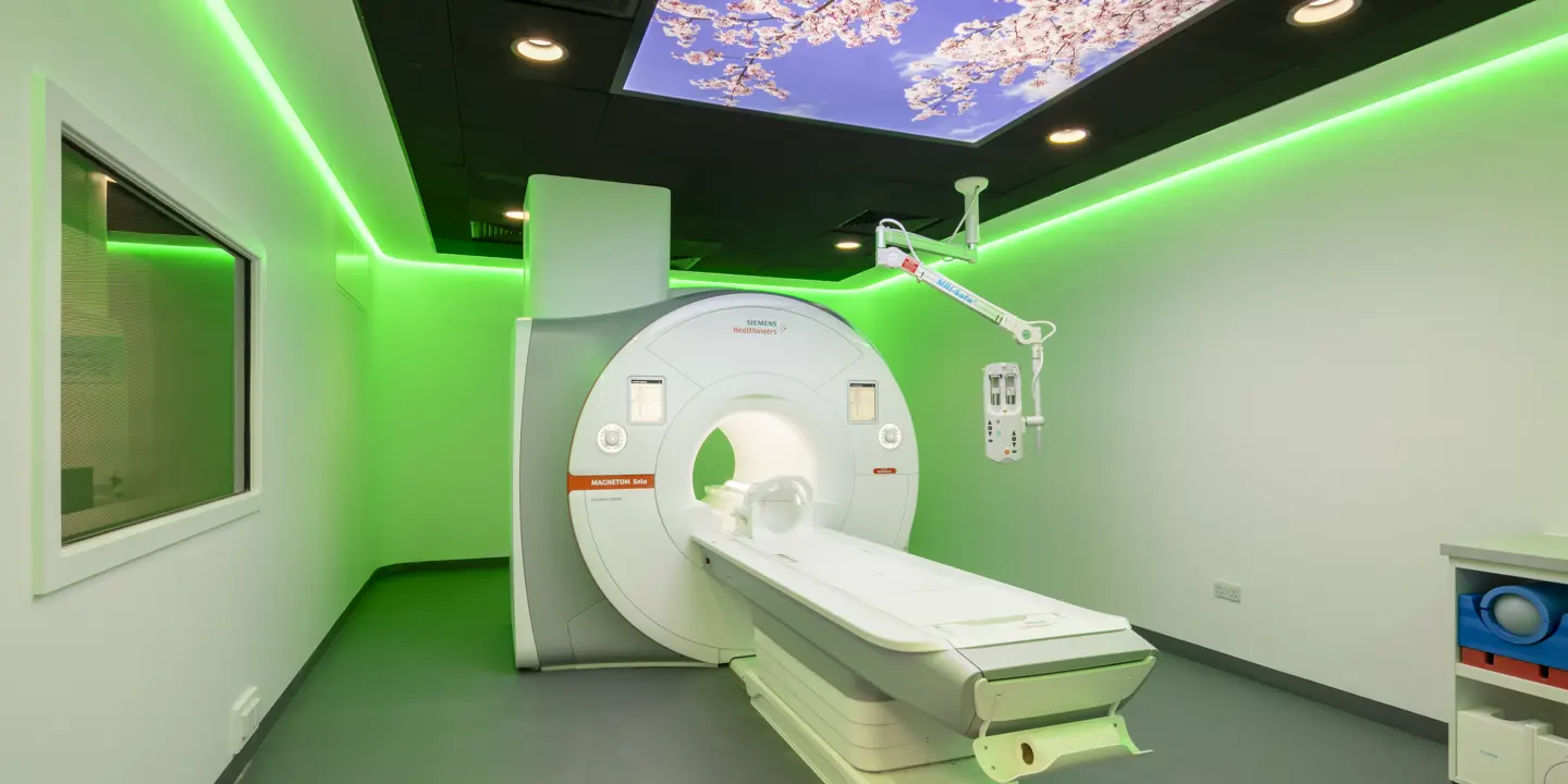
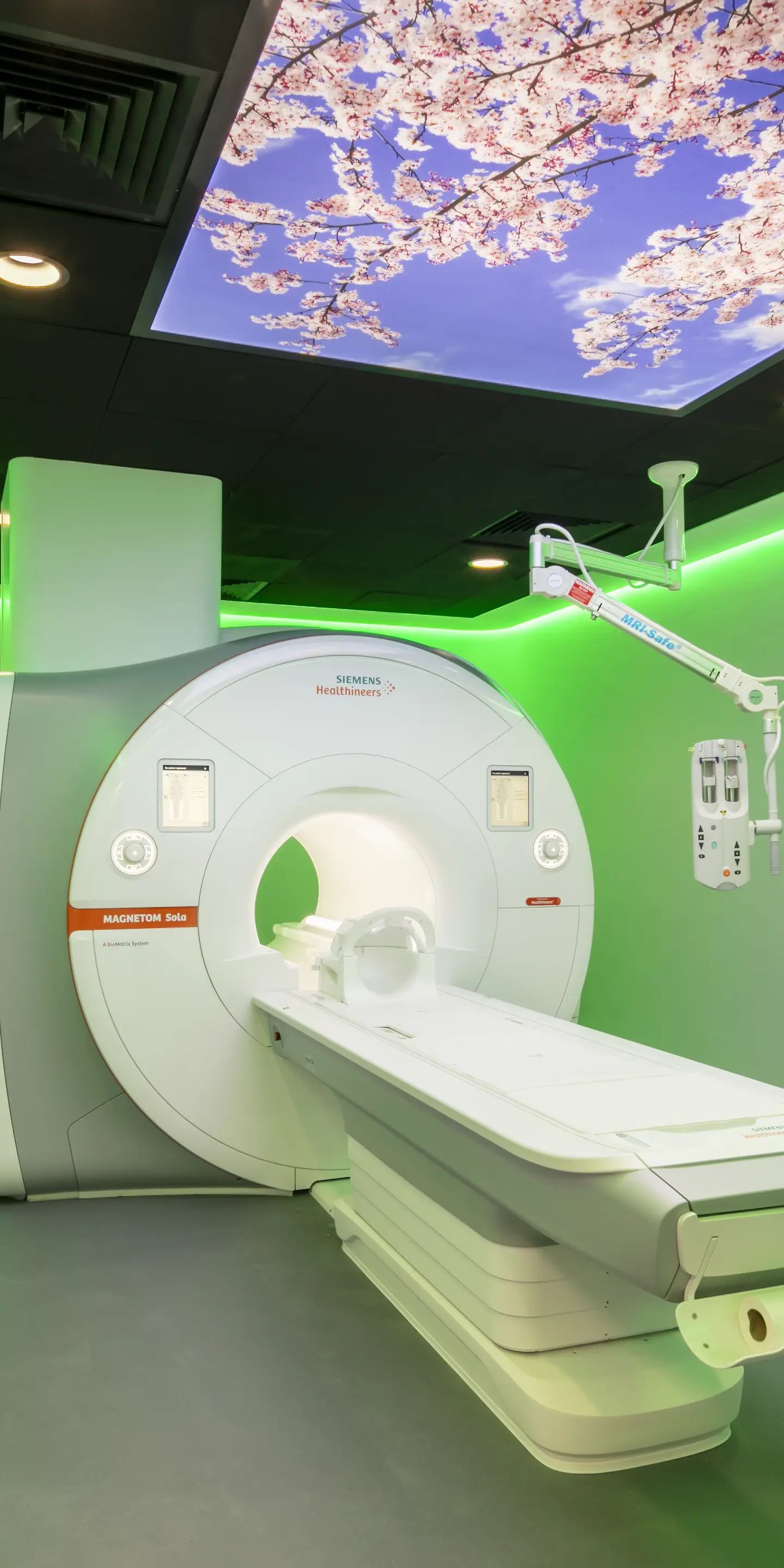
Protons are very sensitive to magnetic fields. During an MRI scan, you will lie back on a scanning table and be slid into a tube-shaped machine. This machine creates a magnetic field around your body using strong magnets. This causes the protons in the hydrogen atoms of the water in your body to line up in the same direction.
Next, short bursts of radio waves are passed through your body. These waves of energy knock the protons out of alignment. When the burst of radio waves stops, the protons once again align with the magnetic field created by the MRI machine. In doing so, they release energy that is detected by sensors in the MRI machine.
How quickly the protons realign with the magnetic field and how much energy is released during this process varies between tissues. Consequently, this creates images that can clearly define the different tissues in your body.
These differences are represented visually by differences in the shades of grey of the MRI images captured. White represents a high-intensity signal, and black represents a low-intensity signal.
The benefits of MRI scans
MRI scans are one of the best ways to detect differences between and within soft tissues. They are particularly useful in detecting abnormal growths, such as those caused by cancer and soft tissue injuries.
This is why people have MRI scans (sometimes referred to as MRI exams). MRIs can help people to investigate unexplained symptoms or be used for general health screening. This supports them to take a proactive approach to their health and well-being.
What’s more, as MRI does not use X-rays, you are not exposed to any ionising radiation from the scan. This means pregnant women in the second and third trimesters can have scans when needed.
MRI scans are not recommended in the first trimester of pregnancy as the radio waves used during the scan can have a heating effect. It isn’t clear what effect this may have on the developing baby at this early point in a pregnancy.
What Can an MRI Scan Detect?
MRI scanners are used to investigate a wide range of symptoms. They can detect diseases and injuries affecting the brain, spinal cord, joints, heart and blood vessels. This includes other organs, such as the kidneys, liver, ovaries, pancreas, prostate, spleen and womb.
Brain MRI scans are often used to diagnose strokes, brain injuries, tumours and aneurysms. They are also used to diagnose degenerative conditions, for example, multiple sclerosis and Alzheimer’s disease.
MRI scans of the spinal cord can help detect compression of the nerves. This can explain symptoms such as numbness and tingling in the limbs and back pain. Spine MRI scans can also detect structural abnormalities present from birth. They can also diagnose structural issues that occur due to ageing, such as degenerative disc disease.
Cardiac MRI scans can help investigate heart problems. For example, structural defects that people may be born with (congenital heart problems). This includes cardiomyopathy, heart valve disease and coronary artery disease.
MRI exams of your joints can detect torn ligaments and cartilage, as well as tumours and bone infections.
MRI exams of other organs can detect a wide variety of conditions. This includes infections, inflammation, cysts, abscesses, fistulas and cancer.
For example, prostate MRI scans can detect several conditions. This includes prostate cancer, an enlarged prostate (benign prostatic hyperplasia) and prostatitis. Meanwhile, gynaecological MRI scans can detect endometriosis, fibroids and womb cancer.
Comparing MRI with Other Imaging Techniques
As mentioned earlier, X-rays and CT scans both expose individuals to ionising radiation. This means that pregnant women are usually advised not to have an X-ray or CT scan. Conversely, MRI scans do not use ionising radiation. This means they are safe to use for pregnant women in the second and third trimesters when needed.
MRI Scans vs CT Scans
Both MRI scans and CT scans (computerised tomography scans) can detect soft tissues, organs and bones. However, the two imaging methods have different strengths. And they are sometimes used in different scenarios.
CT scans can be used for diagnosing the cause of symptoms. They use a combination of X-rays and computer technology to create detailed 3D images of the inside of your body.
As X-rays are a type of ionising radiation, unnecessary exposure to X-rays is not recommended. This is why CT scans are not used for screening, that is, to check your health when you have no symptoms of ill health.
CT scans are better at detecting the margins of one structure versus another. This is known as spatial resolution. Whilst MRI scans are better at detecting differences between tissues and within them. This is known as contrast resolution.
This shows MRI scans are ideal for capturing detailed images. These images can be used to investigate injuries and diseases that affect soft tissues and organs. However, CT scans are faster and are often used in emergency situations. This could be to detect internal bleeding, blood clots and fractures.
Both CT scans and MRI are used to detect cancer and check its progression. However, particular cancers, such as womb cancer, prostate cancer and certain types of liver cancer, are harder to detect using a CT scan.
As MRI does not use ionising radiation, they can be used for screening purposes, unlike CT scans.
MRI Scans vs X-rays
X-rays are most commonly used to investigate injuries and diseases that affect the bones. This includes fractures and structural bone abnormalities (eg scoliosis and bone spurs).
X-rays are a type of ionising radiation that can pass through your body. Your body absorbs X-ray energy at different levels depending on the type of tissue. A nearby sensor detects the X-rays once they have passed through your body. These signals form an image of the inside of your body.
MRI scans can also detect bone injuries and diseases but do not use ionising radiation. They can detect stress fractures in their early stages before they appear on an X-ray.
This is because stress fractures are small cracks that appear in your bones due to repeated bouts of force (stress) being applied to them. An X-ray does not always have the resolution to detect these cracks when they first appear. In contrast, an MRI scan has a very high resolution.
Chest X-rays are used to detect lung cancer, lung infections and chronic (long-term) lung conditions, such as cystic fibrosis and emphysema. Chest MRI scans are less commonly used in these scenarios as they take longer and are more expensive.
MRI scans create detailed 3D images, while X-rays can only capture flat (2D) images.
MRI Scans vs Ultrasound Scans
Both MRI and ultrasound scans do not use ionising radiation. While MRI scans use magnetic fields and radio waves, ultrasound scans use high-frequency sound waves.
Unlike MRI scans, ultrasound scans cannot image hard tissues well, namely bones and joints. They are, therefore, used exclusively to image soft tissues, such as organs, muscles and skin.
Also, while MRI can provide detailed images throughout the full depth of your body, ultrasound is better at capturing detailed images of more superficial structures.
However, an ultrasound scan allows for dynamic imaging. This means your doctor can assess the function of your muscles and tendons as you move them. In contrast, you must remain completely still for an MRI scan.
How MRI Scans Enhance Patient Diagnosis
MRI scanners have the ability to detect subtle differences between different types of tissues. They can show the contrast between healthy and abnormal tissues, which means they can aid in the early diagnosis of several conditions.
This is particularly vital when diagnosing cancer and monitoring its progress. MRI scans are often the preferred imaging tool for investigating symptoms of brain and spinal cord tumours.
When it comes to symptoms of prostate problems, which could be caused by prostate cancer, an MRI scan can help your doctor reach a diagnosis without a more invasive and uncomfortable rectal examination. Prostate MRI scans can also form part of a general health assessment so you can take a preventative approach to your prostate health.
Similarly, breast MRI scans can be used as part of a routine breast health check-up to catch the early signs of breast cancer. Breast MRI scans can detect smaller tumours that may be missed with a standard mammogram. Therefore, breast MRI scans can detect breast cancer at an earlier stage.
Breast MRI scans are sometimes recommended for younger women who tend to have more dense breast tissue and women with breast implants. They are also offered annually via the NHS to women aged 25 and above who have certain genetic flaws (mutations) in their BRCA1 or BRCA2 genes as these mutations dramatically increase the risk of breast cancer.
Common Concerns about MRI Scans
One of the most common concerns about MRI scans is claustrophobia since it takes place inside a tube-shaped machine. The machine also makes loud banging, clicking and whirring sounds, which can make some individuals feel anxious. If you are concerned about how you will feel during your MRI scan, it is important to tell your care team.
At Vista Health, we will do our best to make you feel as comfortable as possible. MRI machines allow for two-way communication. This means our radiographers can speak to you during your scan and you can let them know if you need to pause or stop the scan.
Another common concern about MRI scans is the use of a gadolinium contrast agent. This is a rare earth metal that is injected into a vein and alters the magnetic properties of water molecules in your body. This helps create greater contrast and so clearer images.
Gadolinium is generally tolerated well. However, in rare cases, individuals can have an allergic reaction to gadolinium. This is why if you have an MRI scan with gadolinium, you need to remain at the scanning facility for 30 minutes after your scan. This is in case you develop an allergic reaction.
If you have previously had an allergic reaction to a different contrast agent, such as an iodine-based contrast agent (used during a CT scan), it does not mean you will develop an allergic reaction to gadolinium. However, it is important to inform your care team of your previous allergic reaction.
Discussing MRI Options with Your Doctor
If you have unexplained symptoms that are a concern, you can see your GP. They will ask you about your symptoms and medical history and then determine whether or not an MRI scan would be appropriate.
There are 2 main types of MRI scans for diagnosing conditions: T1 and T2. The differences between these 2 scans are due to different timings of the pulses of radio waves that are used to create MRI images.
T1 MRI scans capture clearer images of fat and are used to detect tumours and structural abnormalities. T2 MRI scans capture clearer images of fluids. This means they are useful in detecting inflammation, infection and swelling.
If you need to have an MRI scan, your doctor will determine which type of scan is needed.
Do You Need a Referral for an MRI Scan?
You do not need a referral from your GP or other healthcare professional to have a private MRI scan in the UK. At Vista Health, you can self-refer and book an MRI scan at one of our nationwide clinics.
We offer both full-body MRI scans and targeted MRI scans such as prostate, cardiac and gynaecological MRI scans. MRI scans of other body parts and organs are also available, including for the head, spine, bowel, chest and limbs.
Whether you would like to investigate any concerning symptoms or want the peace of mind that comes with a full-body MRI scan, we can help. Book your private MRI scan with Vista Health today.
Sources
https://www.rbht.nhs.uk/blog/history-magnetic-resonance-imaging-mri
https://www.nibib.nih.gov/science-education/science-topics/magnetic-resonance-imaging-mri
https://www.fda.gov/radiation-emitting-products/mri-magnetic-resonance-imaging/benefits-and-risks
https://www.mayoclinic.org/tests-procedures/mri/about/pac-20384768
https://www.mdanderson.org/cancerwise/ct-scan-vs-mri--what-is-the-difference.h00-159616278.html
https://www.mayoclinic.org/diseases-conditions/stress-fractures/diagnosis-treatment/drc-20354063
https://www.hopkinsmedicine.org/health/treatment-tests-and-therapies/magnetic-resonance-imaging-mri
https://www.hopkinsmedicine.org/health/treatment-tests-and-therapies/breast-mri
https://www.guysandstthomas.nhs.uk/health-information/gadolinium-contrast-injection