Benefits of Ultrasound: Discover Key Advantages for Health
Ultrasound is an affordable, painless and non-invasive medical imaging technology. It is also known as sonography or ultrasonography. It uses sound waves to create images of internal organs, tissues, and other structures inside a human body.
But what are the advantages of ultrasound scans for your health? Here, we’ll explore ultrasound benefits. Read on to learn how it works, which medical conditions it can help to diagnose and treat, and what you can expect from your ultrasound scan.
What is ultrasound?
Unlike other imaging methods, such as a CT scan or X-ray, ultrasound technology does not use ionising radiation. Instead, it uses high-frequency sound waves to create images of the inside of your body. You will not be able to hear these sound waves as they are beyond the limits of human hearing. The name ‘ultrasound’ comes from the high frequency of these sound waves.
During an ultrasound scan, the ultrasound waves will pass into your body. They will be reflected back when they meet a boundary between different types of tissues. For example, between soft tissues and bone or soft tissues and fluid. These reflected ultrasound waves are then detected by the ultrasound imaging machine and converted into electrical signals.
The electrical signals are turned into grayscale images. Each point of brightness on the image represents an electrical signal. On average, more than 1,000 high-frequency sound waves can be reflected back in just one second, so images of the inside of your body can be generated in real-time.
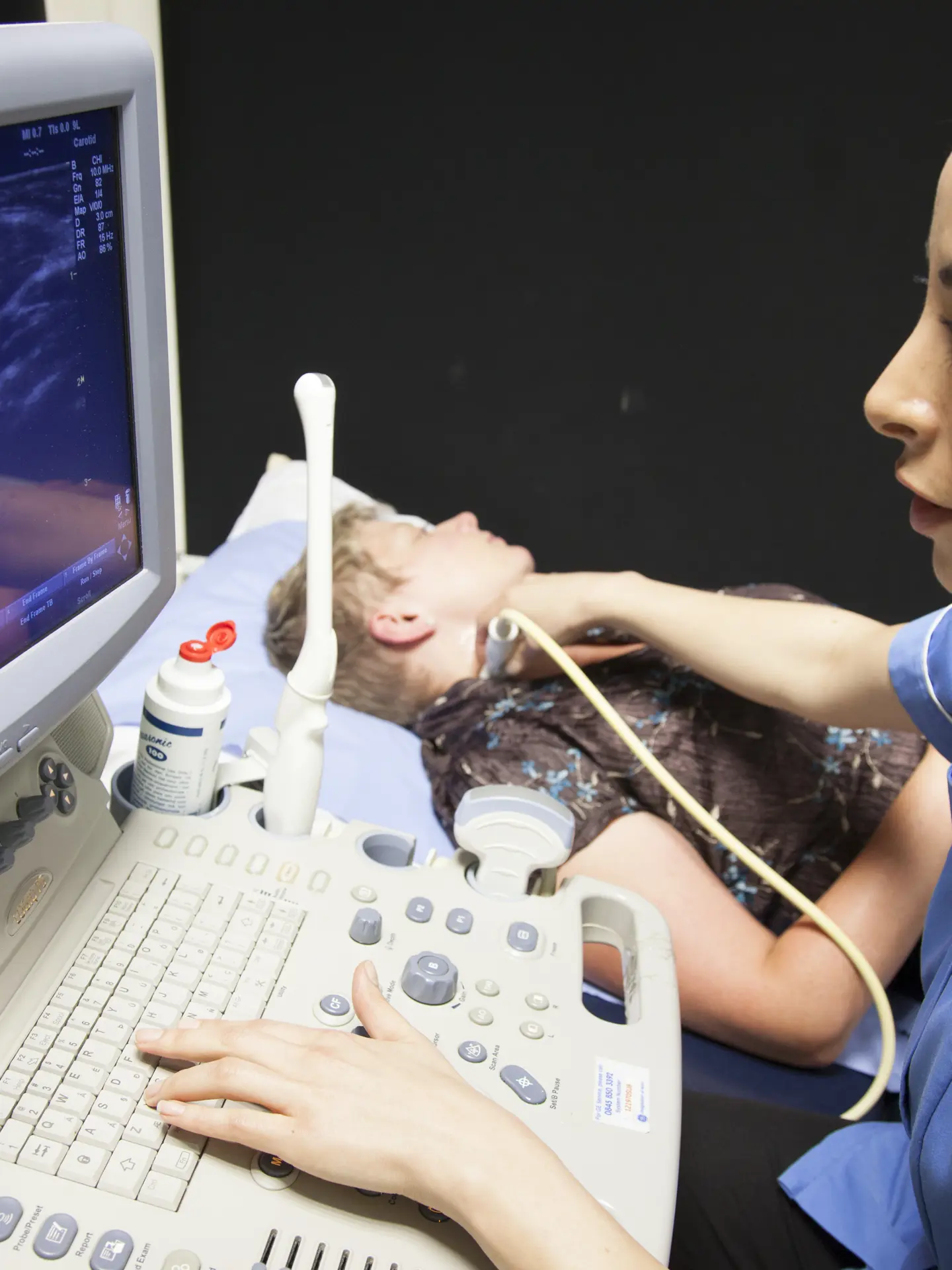
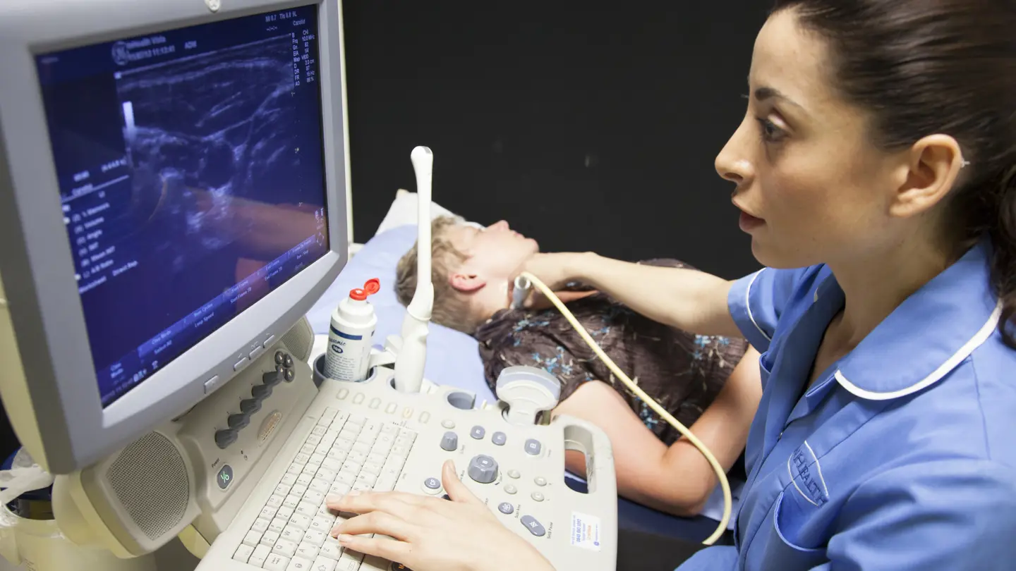
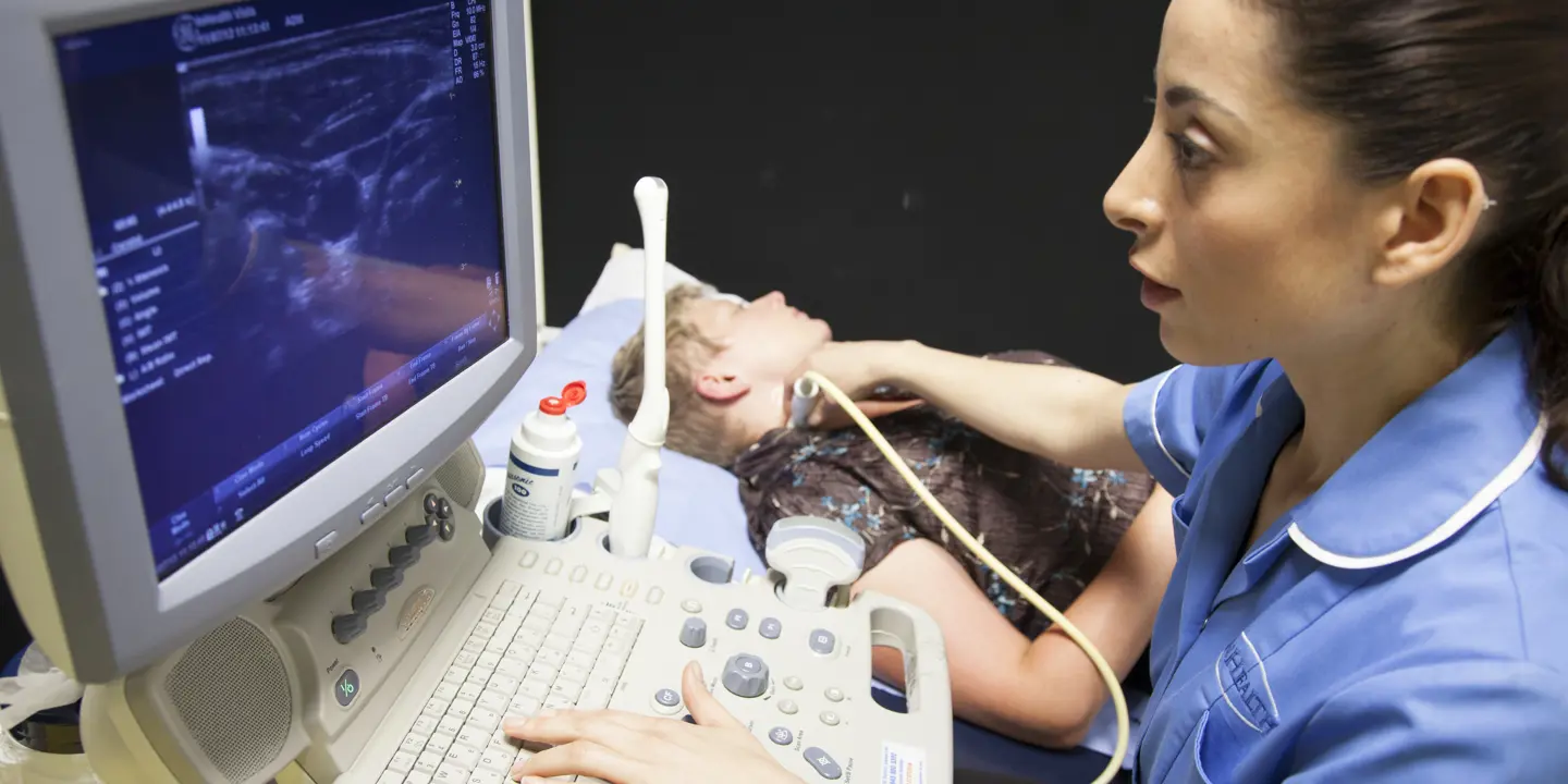
What does the ultrasound procedure involve?
Ultrasound machines are much smaller than CT or MRI machines. Therefore, they can be moved around. The machine includes a computer console, digital screen and transducer. The transducer is a handheld probe that comes in different shapes and sizes depending on which part of the body needs to be imaged.
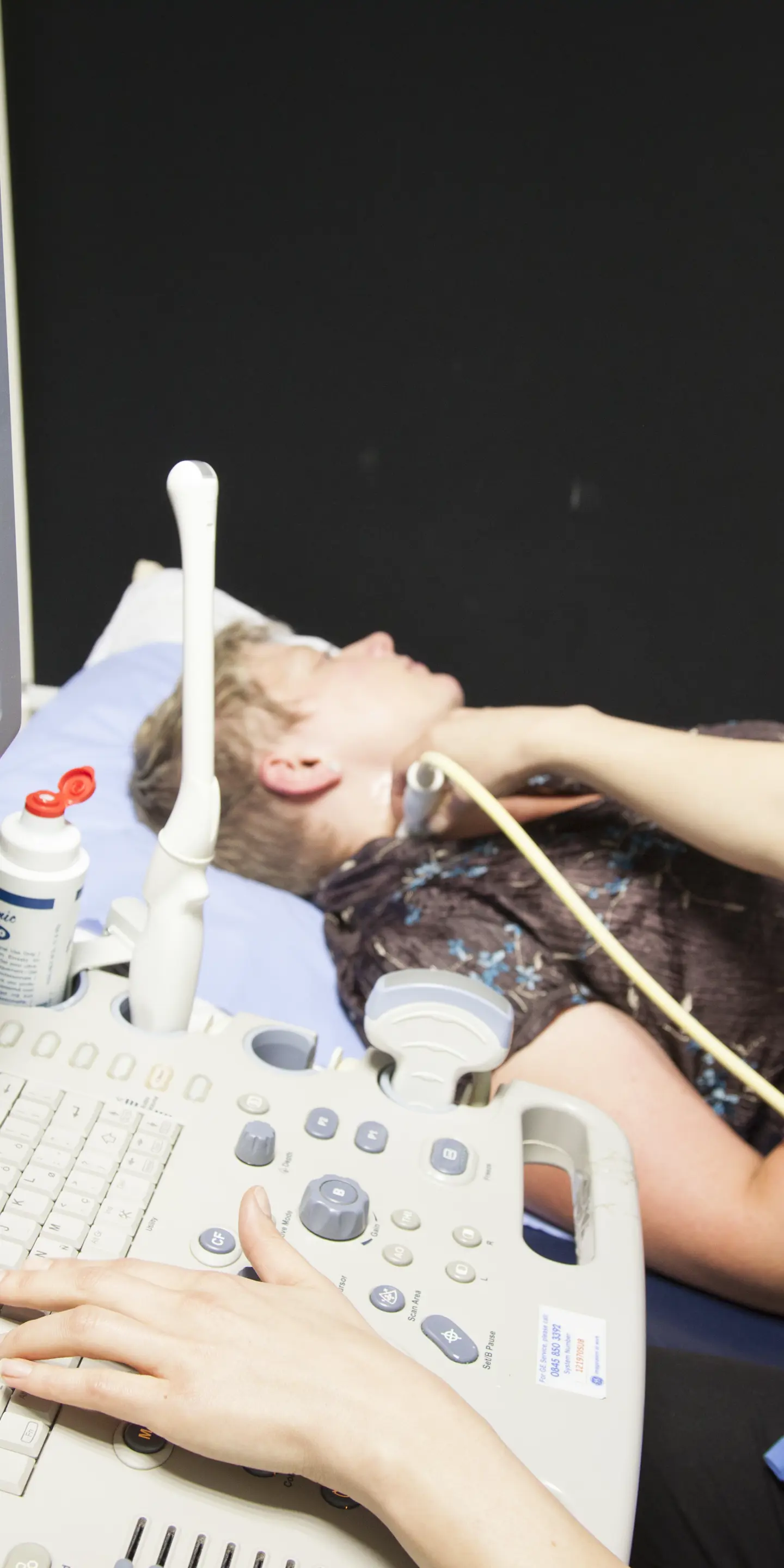
During your ultrasound scan, you will be asked to lie back on an examination table. Depending on which part of your body is being scanned, you may be asked to lie completely flat, sitting partially upright or turned onto one side of your body.
The area of skin that’s being scanned will be cleaned. As the scan uses sound waves, rather than light waves, body hair will not interfere with the scan and does not need to be shaved off. However, you may be asked to change into a hospital gown or move your clothing out of the way depending on which area of your body is being scanned.
A water-based gel will be applied to your body. The ultrasound gel improves the contact between the transducer and your body. It also reduces the chances of pockets of air coming between the transducer and your body. Air pockets can block sound waves from travelling and interfere with the scan.
The transducer will be placed along the part of your body that’s being imaged. It will release sound waves into your body as it is moved along it. The transducer will also detect the reflected sound waves. It may be tilted at different angles to capture a better and/or more complete view of the inside of your body.
The transducer will convert these reflected sound waves into electrical information, which will be sent to the computer console. The computer console will convert the electrical information into visual information, which will be displayed on-screen in real-time.
At Vista Health, we aim to deliver the results of your scan within 3 working days. Your report will provide a summary of your scan results and insights from one of our experienced radiologists. A radiologist is a doctor who specialises in diagnosing and treating conditions using medical imaging.
Your Vista Health ultrasound report can be sent to your NHS GP or you can book a private GP appointment with one of our expert GPs.
Understanding the benefits of ultrasound
Ultrasound is non-invasive
During an ultrasound scan, no cuts will be made into your body – unlike some keyhole investigations like laparoscopy (to investigate the pelvis or abdomen) and thoracoscopy (to investigate the chest).
Ultrasound is painless so you don’t need any general or local anaesthesia.
Ultrasound works in real-time
While ultrasound scans don’t provide the same level of detail as a CT scan or MRI scan, they generate images of the inside of your body in real time. This is thanks to the speed that sound waves are reflected back by tissues in your body.
This means ultrasound scans can be used for dynamic imaging ie where you can move part of your body to see how your internal structures respond in real time. This is useful for investigating musculoskeletal problems.
Ultrasound is cost-effective
An ultrasound does not use ionising radiation or powerful magnets, but instead uses sound waves. This technology is less expensive to make and maintain, so the scan is cheaper compared to CT scans and MRI scans.
Who can have an ultrasound scan?
Ultrasound imaging does not create radiation exposure, as it does not use ionising radiation. So, ultrasounds are safe for use in children and pregnant women for foetal imaging. Ultrasound scans are routinely used to check foetal development and the health of a growing baby during pregnancy.
As ultrasound scans do not involve the powerful magnets that are used in MRI scans, they are safe for people with metal implants or fragments in their bodies.
The ultrasound machine is small. People who have claustrophobia don’t need to worry about feeling enclosed or trapped during the scan, which can happen during an MRI scan or CT scan.
All of this means that most people can safely have an ultrasound scan.
Which conditions can ultrasound help diagnose?
Ultrasound can be used in many ways to diagnose conditions affecting multiple organs and tissues. That means a diagnostic ultrasound can detect disease and tissue damage early so it can be treated before it gets worse. For example, ultrasound can pick up the early signs of liver disease, cardiovascular conditions and fertility problems.
Abdominal ultrasound
Ultrasound scans of the abdomen are often used to investigate conditions affecting abdominal organs such as the kidneys and liver. This includes:
-
liver lesions that may be benign (non-cancerous) or cancerous
-
fatty liver disease
-
kidney cancer
-
kidney cysts
-
kidney stones
-
kidney infections.
An abdominal ultrasound can also be used to investigate the pancreas and gallbladder to help diagnose inflammation of the pancreas (pancreatitis), pancreatic cancer, gallstones and inflammation of the gallbladder (cholecystitis).
When the bladder is full, it extends from the pelvis into the abdomen. Ultrasound scans of the bladder can detect bladder stones, bladder cancer and an overactive bladder.
Pelvic ultrasound
A pelvic ultrasound scan in women often involves the use of a transvaginal transducer or probe that is inserted into the vagina.
This can help diagnose a variety of gynaecological conditions, including pelvic inflammatory disease (PID), ectopic pregnancy, ovarian cysts, uterine fibroids and womb cancer.
Pelvic ultrasound can also help detect irregularities within the pelvic organs, for example, whether the womb is shaped abnormally. A unicornuate (one-sided), bicornuate (heart-shaped) or septate (divided) womb can have a negative impact on fertility.
Women may have a pelvic ultrasound to investigate their fertility status by checking the number of follicles in the ovaries. A follicle count gives an idea of how many eggs are left in the ovaries, which is used to predict fertility levels.
A pelvic ultrasound scan cannot definitively diagnose endometriosis, but it can be used as a step on the way to confirming a diagnosis.
In men, a pelvic ultrasound scan can investigate the health of the prostate and testicles. A transrectal probe is inserted into the rectum. It can be used to help diagnose an enlarged prostate, prostate cancer and inflammation of the prostate (prostatitis).
Pelvic ultrasound of the testicles can help detect testicular cysts and cancer, twisting of the cord that supplies blood to the testicles (testicular torsion) and enlargement of the veins of the scrotum (varicoceles).
Cardiovascular ultrasound (echocardiogram)
Ultrasound is used to image the structure of the heart, including its chambers and valves, as well as to image its blood supply. This is called an echocardiogram and can help assess the health and function of the heart, as well as evaluate blood flow.
An echocardiogram can help diagnose:
-
heart failure
-
congenital (from birth) heart defects
-
thickening of the heart walls (cardiomyopathy)
-
infection of the heart lining (endocarditis)
-
heart valve problems
-
tissue damage after a heart attack.
There are two main types of echocardiogram: transthoracic echocardiogram (TTE) and transoesophageal echocardiogram (TOE).
TTE is non-invasive as the ultrasound transducer is passed across your chest. It is the most common type of echocardiogram. It can be used during an exercise stress echocardiogram, where images of the heart are captured before and after you exercise on a treadmill. This shows how your heart responds to stress.
Most ultrasounds are non-invasive procedures. TOE is invasive. A thin, flexible telescope-like device with a camera and light on the end (endoscope) is passed down your throat and into your gullet (oesophagus). The endoscope provides live video images of the inside of your body. This allows the ultrasound transducer to be safely guided down into your oesophagus.
TOE may be recommended if the ultrasound images captured during a TTE were not clear enough.
Ultrasound for musculoskeletal health
Ultrasounds are a great diagnostic tool for musculoskeletal chronic pain. An ultrasound exam is often used to investigate musculoskeletal problems, such as joint pain and swelling. General ultrasound imaging can be used dynamically to find out how movement affects the tissues in a joint.
This can be helpful to diagnose conditions affecting the bursae, such as inflammation of the bursae (bursitis). Bursae are fluid-filled sacs in the joints that reduce friction between joint bones so they can move smoothly.
Inflammation of the tendons (tendonitis) and tears can also be detected via ultrasound. Common tendon injuries include:
-
rotator cuff tears of the shoulder joint
-
Achilles tendon ruptures of the ankle
-
patellar tendon tears of the knee
-
bicep tendon tears of the upper arm.
Ultrasound of the ligaments can help detect sprains and tears too. Common ligament injuries include:
-
anterior cruciate ligament tears of the knee
-
lateral ligament sprains of the ankle
-
iliotibial band syndrome ie inflammation of the tendon running along the outer side of the body from the hip to the knee.
Ultrasound can also diagnose:
-
fluid-filled sacs called ganglion cysts that are found near joints
-
muscle tears, growths and oedema (fluid build-up)
-
the early signs of inflammatory arthritis.
Ultrasound-guided procedures
Ultrasound is not only used to investigate symptoms and diagnose conditions. It can also be used in the treatment of conditions.
This includes steroid joint injections, the removal of fluid from cysts (cyst aspiration), and treatments for carpal tunnel syndrome and trigger finger.
Explore ultrasound options for your health
If you have unexplained symptoms, such as pain, discomfort, swelling or tenderness, an ultrasound scan can help get to the bottom of it.
At Vista Health, you can access advanced ultrasound scanning if you think you have an injury or you’re concerned about potential disease. You do not need a referral from a doctor. You can self-refer online and book your private ultrasound at one of our conveniently located scanning facilities nationwide.
Sources
https://www.mayoclinic.org/tests-procedures/ultrasound/about/pac-20395177
https://my.clevelandclinic.org/health/diagnostics/4995-ultrasound
https://www.medicalnewstoday.com/articles/245491
https://www.nhs.uk/conditions/ultrasound-scan/
https://www.guysandstthomas.nhs.uk/our-services/ultrasound/appointments
https://www.sciencedirect.com/science/article/abs/pii/S0169409X2400022X
https://pmc.ncbi.nlm.nih.gov/articles/PMC7602142/
https://my.clevelandclinic.org/health/diagnostics/4994-abdominal-ultrasound
https://pmc.ncbi.nlm.nih.gov/articles/PMC5894446/
https://www.nhs.uk/conditions/endometriosis/
https://www.hopkinsmedicine.org/health/treatment-tests-and-therapies/pelvic-ultrasound
https://www.radiologyinfo.org/en/info/us-prostate
https://www.radiologyinfo.org/en/info/us-scrotal
https://my.clevelandclinic.org/health/diagnostics/13477-echocardiogram-transthoracic-tte
https://my.clevelandclinic.org/health/diagnostics/16947-echocardiogram