CT Scans: Can They Detect Lung Problems Effectively?
CT scans are used to create images of the inside of your body to detect a range of health conditions. This includes problems in the lungs. So, why would a doctor order a CT scan of the lungs?
Put simply, a CT scan can help to investigate the possible cause of lung symptoms such as a persistent unexplained cough, breathlessness and/or chest pain.
You can also have a private lung CT scan as part of a Lung Health Assessment if you are concerned about a family history of lung problems. This can empower you to take the right steps to reduce your risk of lung disease.
Here, we will explore how CT scans identify lung issues and common lung conditions they can detect. We will also discuss the risks and benefits of lung CT scans.
What is a CT scan?
CT stands for computed tomography. Tomography is a technique for displaying a cross-sectional 3D image through an object. In the case of a CT scan, a 3D image is created from layers of cross-sectional images stacked on top of each other. These are generated using X-rays and computer technology.
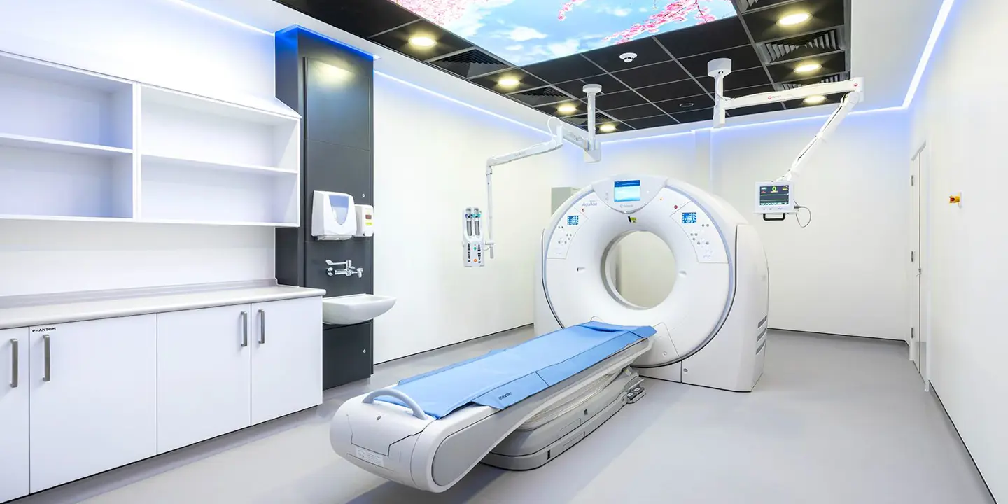
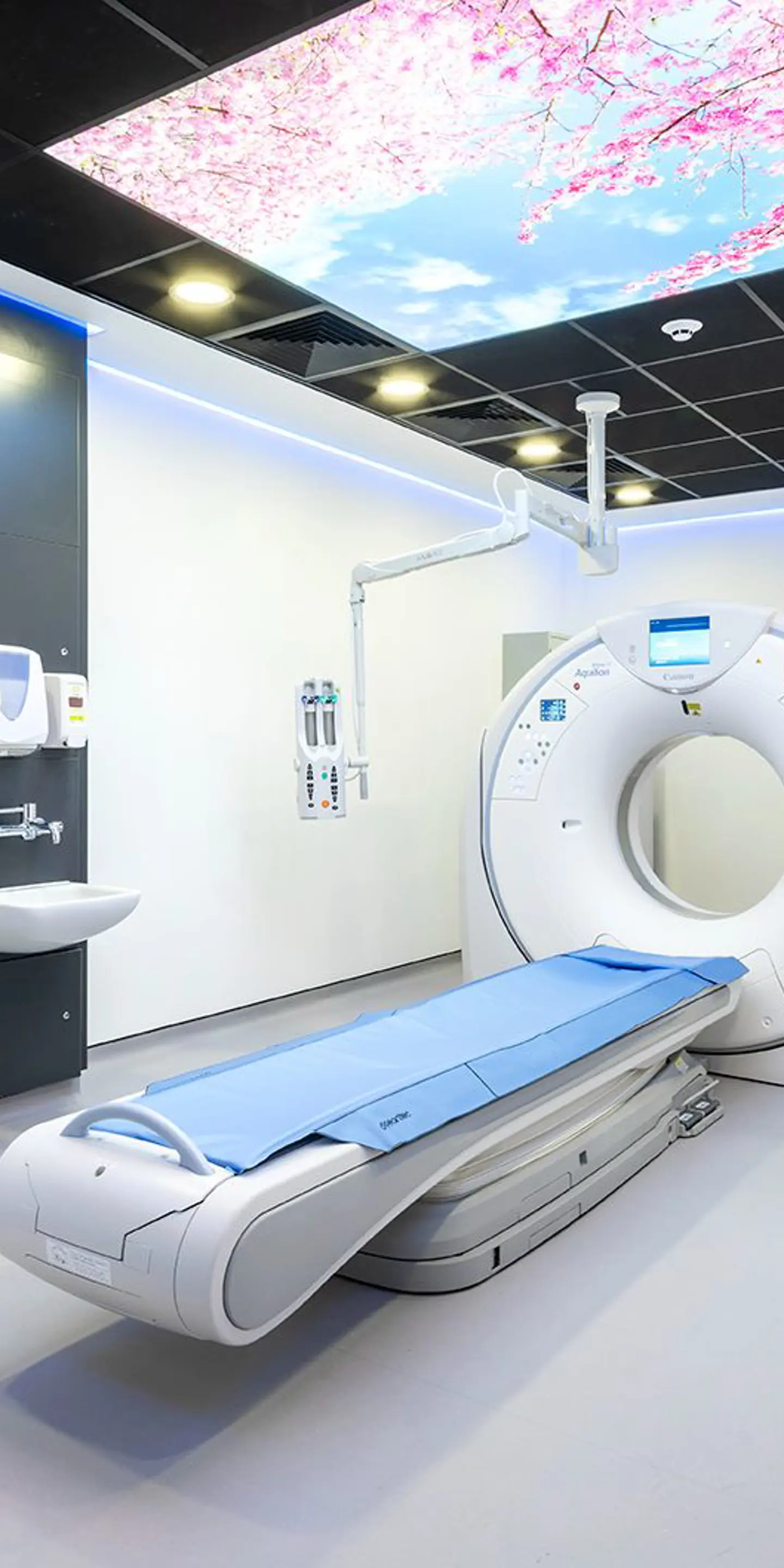
A CT scanner is made up of a ring-shaped structure, an examination table and a computer. During your scan, you will lie back on the examination table. It will then be passed through the ring-shaped structure, which will rotate around your body.
As the ring rotates around you, it will release narrow beams of X-rays that will pass through your body. The ring has sensors which will detect the X-rays once they have passed through your body, and send signals to the computer for processing.
As different tissues absorb different amounts of X-rays, a CT scan can detect a variety of tissues and structures. This includes your blood vessels, bones, cartilage, muscles, organs and soft tissues (e.g. ligaments and tendons).
The computer will compile the information it received from the X-ray detectors to create cross-sectional images through your body. These images can be viewed as slices, or stitched together to form a 3D image of the skeleton, body tissues, organs, and any abnormalities in the area being scanned.
A lung CT scan can detect changes in your lung tissue, the blood vessels that supply your lungs, the membranes that cover your lungs (pleural membranes) and the airways of your lungs (bronchi and bronchioles).
Contrast-enhanced CT scans
In certain cases, your doctor may recommend that you have a contrast-enhanced CT scan. This involves injecting an iodine-based contrast agent (a special dye) into a vein in your arm. The dye itself is clear and does not stain your organs.
It helps create clearer images during your scan as it absorbs more X-rays. Areas that absorb more X-rays appear brighter on the CT scan. Consequently, areas with high levels of contrast agent will be highlighted on the scan. For a lung CT scan, this means blood vessels supplying the lungs appear more clearly.
Benefits of a lung CT scan
Early detection of disease
The lungs are large organs, and when damaged, they have a great capacity to compensate for that damage. This means that many lung conditions only cause symptoms when the disease is quite advanced.
As with any disease, the more advanced it is, the harder it is to treat. A lung CT scan can help detect negative changes in your lung tissue before you notice any symptoms. Early detection of lung disease means you can seek treatment sooner to avoid developing symptoms.
This can be particularly helpful if you have a family history of lung cancer, familial pulmonary fibrosis or other conditions where genetics may play a role. In the 2016 UK Lung Cancer Screening Trial, approximately 85% of the lung cancers detected were at an early stage thanks to preventative CT scans.
High resolution
Unlike a chest X-ray, which produces 2D images, a lung CT scan produces detailed 3D images. These images are created at a higher resolution than X-rays. This means they can detect more subtle changes in your lungs than a chest X-ray and can better determine what that change suggests about your lung health.
For example, a chest X-ray may detect a mass in your lungs but it may not be able to determine whether it is a tumour or an abscess (a pus-filled mass). A lung CT scan can detect the difference in these tissue types, which means you can get the right treatment sooner.
Rapid imaging
A CT scan of your lungs usually takes 10–30 minutes. Although this is longer than a chest X-ray, which only takes a few minutes, the images created are much more detailed. What’s more, this is much quicker than a chest MRI scan, which can take up to 90 minutes.

Risks and limitations of a lung CT scan
Exposure to radiation
X-ray technology uses ionising radiation. Consequently, both CT scans and X-rays expose you to ionising radiation. Ionising radiation can damage DNA, and while your cells can repair this damage, there’s a risk of damage being repaired incorrectly. This can lead to mutations in your DNA that may increase your likelihood of developing cancer in the future.
A CT scan of your lungs exposes you to more radiation than a chest X-ray. However, the levels of radiation used in a chest CT scan are still considered to be low.
For context, over a year, an individual in the UK is, on average, naturally exposed to 2.7 millisieverts (mSv) of radiation from the environment. A standard chest CT scan exposes you to around 6.6 mSv, while a chest X-ray exposes you to around 0.02 mSv.
At Vista Health, we take advantage of the latest advances in CT imaging. This means we can offer our customers low-dose CT scans of the lungs as part of our Lung Health Assessments.
A low-dose CT scan of the lungs exposes you to up to 90% less radiation than a standard CT scan of the lungs, but still produces detailed images.
Detection of incidental findings
A lung CT scan can pick up both cancerous and non-cancerous growths. However, it may not be clear from your initial lung CT scan whether a lung nodule is cancerous or not.
In most cases, lung nodules are not cancerous. They are often the result of a previous lung infection and are not a cause for concern. Such findings after a health check are often called incidental findings.
Once a lung nodule has been detected, you will most likely need a follow-up lung CT scan several months later to rule out lung cancer ie to check that the nodule is not growing. This can potentially cause unnecessary worry while waiting for your next scan results.
Reaction to the contrast agent
If you need to have a contrast agent for your lung CT scan, there is a very small risk of developing an allergic reaction.
Up to one in every 100 people develop a mild allergic reaction to the contrast agent used in CT scans. Symptoms include the following:
-
a mild rash
-
flushing
-
headaches
-
itchiness
-
nausea and/or vomiting
-
swelling
Symptoms of a mild to moderate reaction usually resolve on their own without any medical intervention. However, you should still inform your care team as soon as possible, so you can be monitored in case your reaction becomes more severe.
A severe reaction to a contrast agent is even more rare than mild to moderate reactions. It usually occurs within 20 minutes of having the contrast agent. This is why you will be asked to remain at the scanning facility for up to 30 minutes for monitoring after your contrast-enhanced CT scan.
When doctors recommend lung CT scans
Your doctor may recommend a lung CT scan if you have any of the following symptoms:
-
a persistent cough — as most coughs get better within three weeks, a persistent cough is defined as one that lasts three or more weeks
-
breathlessness — this may be on exertion (eg when walking around or walking up stairs) or at rest
-
chest pain
-
fever
Whether a CT scan is the most appropriate scan for you will depend on several other factors. This includes your medical history, other associated symptoms and the results of any other tests you have had.
Common lung conditions detected by CT scans
A CT scan of your lungs can help diagnose a variety of lung conditions, including the following:
A chest infection
Infections of the lungs can be caused by many different microorganisms, including bacteria (eg tuberculosis and Streptococcus pneumoniae), viruses (eg influenza and COVID-19) and fungi (eg Aspergillus).
Bronchiectasis
This refers to widening of the airways (bronchi) of the lungs over a long period of time. It is often caused by previous infections of the lungs that damage the bronchi.
Emphysema
This refers to enlargement of the air sacs that comprise your lung tissue (alveoli). The most common cause of emphysema is chronic (ie long-term) obstructive pulmonary disease (COPD), which is most often caused by smoking.
Interstitial lung disease (ILD)
This refers to a group of over 200 different conditions that damage the interstitium of the lungs. The interstitium comprises the connective tissue that fills the space between the alveoli of your lungs and the blood vessels around these alveoli.
When ILD damages the interstitium, it can cause inflammation and in some cases thickening and scarring (fibrosis). Examples of ILD include sarcoidosis, idiopathic pulmonary fibrosis (IPF) and hypersensitivity pneumonitis.
Lung cancer
This refers to the uncontrolled growth of cells that originate in the lungs. It is the third most common cancer in the UK and can be divided into two types: small-cell lung cancer and non-small-cell lung cancer. It’s classified by the type of cells the cancer is found in and each type may require different treatment. Small-cell lung cancer usually spreads faster, but is less common than non-small-cell lung cancer. The majority (79%) of lung cancer cases in the UK are preventable with lifestyle changes to avoid common risk factors, such as quitting smoking.
Your CT scan procedure
If you are having a contrast agent for your lung CT scan, you will be asked not to eat or drink anything other than water for up to four hours before your scan.
On the day of your scan, wear loose, comfortable clothing and remove any metal accessories or jewellery around the chest area.
If you are having a contrast agent, this will be injected into a vein in your arm before you have your scan.
During your scan, you will be asked to lie back on the examination table while the ring of the CT scanner rotates around you. At certain points, your radiographer may ask you to momentarily hold your breath — this can help create clearer images by reducing blurring caused by movement.
After your scan, you will be asked to remain at the scanning facility for up to 30 minutes if you have had a contrast agent. If not, you will be able to return to your usual activities straight away.
At Vista Health, we aim to deliver a report with your results and insights from one of our experienced radiologists within 3 working days.
Your results can be sent to your NHS GP or you can book an appointment with one of our expert GPs to discuss your results. They will explain exactly what they mean for your particular circumstances and advise you on any next steps depending on the findings.
Find out where you stand with your lung health
If you’re concerned about unexplained lung symptoms or are worried about a family history of poor lung health, Vista Health can help.
Get expert insights into your lung health today when you book a private CT scan of your lungs at any one of our nationwide clinics.
Sources
https://www.radiologyinfo.org/en/info/chestct
https://www.radiologyinfo.org/en/info/safety-xray
https://www.radiologyinfo.org/en/info/screening-lung
https://www.nhs.uk/conditions/lung-cancer/diagnosis/
https://www.healthline.com/health/how-long-does-an-mri-take
https://www.cancer.org/cancer/types/lung-cancer/detection-diagnosis-staging/lung-nodules.html
https://www.dgft.nhs.uk/wp-content/uploads/2023/08/CT-and-MRI-scan-post-reaction-V1.pdf