
Head MRI Scan: What It Can Detect and Diagnose
A head MRI scan (sometimes called a brain MRI or cranial MRI) captures highly detailed 3D images of the inside of your head. Brain MRI scans can help detect early signs of disease, such as brain tumours, and brain damage, such as that caused by a stroke or traumatic brain injury.
A head MRI scan can also uncover the cause of unexplained symptoms, such as changes in your behaviour or persistent headaches.
We’re often asked ‘What can a head MRI scan detect?’. Join us below as we uncover the answer. First, it helps to understand more about what an MRI scan involves.
What is MRI?
MRI or magnetic resonance imaging takes advantage of the magnetic properties of water molecules in your body to create the most detailed pictures possible of any medical imaging tool today.
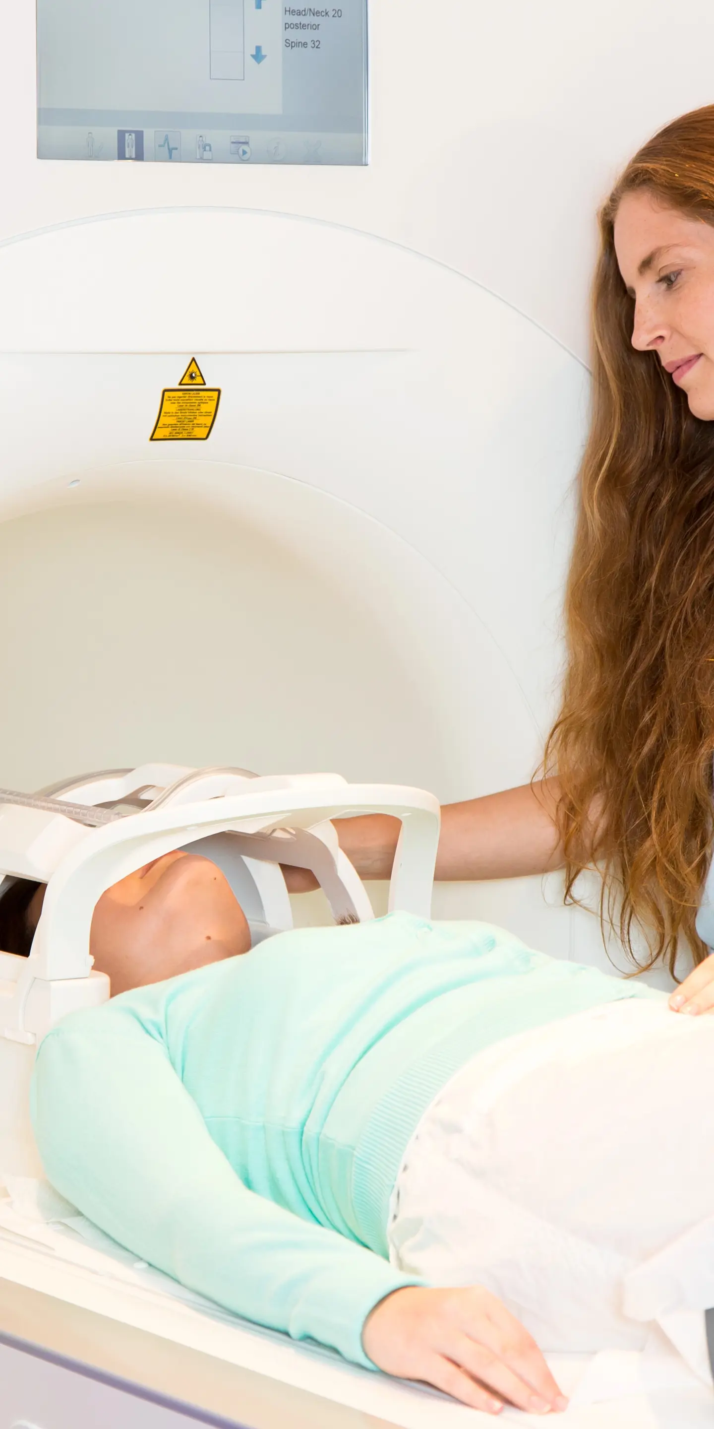
It uses powerful magnets that form the tube structure of the MRI machine. During your MRI scan (sometimes called an MRI exam), you will lie back on a table that slides into the tube of the MRI machine. The magnets then create a magnetic field around your body.
This magnetic field affects the hydrogen atoms that comprise the water molecules in your body. Specifically, it affects tiny particles called protons in the hydrogen atoms and causes them to all point in the same direction.
Then, the MRI unit releases pulses of radio waves that pass through your body. These radio waves push the protons out of alignment. When the radio waves stop, the protons realign. This process releases energy in the form of radio signals that are detected by the MRI machine.
The radio signals are converted into 3D images of the inside of your body. The images are white, black and varying shades of grey.
Areas of your body with a high water content, such as fat or cerebrospinal fluid in the brain, produce high-intensity signals and, therefore, appear white. Areas of your body with a low water content, such as hard bone or air, produce low-intensity signals and, therefore, appear black.
Understanding head MRI scans
A head MRI scan provides high-resolution images of the structures and tissues inside your head. This includes your:
-
brain and its meninges (the three layers of tissue that cover your brain)
-
cerebral blood supply, that is, all the blood vessels that supply your brain
-
skull and facial bones
-
eyes and inner ear
-
fat, muscle and connective tissue
-
nerves supplying your head.
When you have a head MRI scan, your whole body will be slid head-first into the tube of the MRI machine. The MRI scanner usually takes 30–90 minutes.
Common conditions detected by head MRI
Common conditions that can be detected via head MRI imaging tests include:
-
brain tumours, which may be cancerous or non-cancerous (benign)
-
neurodegenerative conditions eg Alzheimer’s disease and multiple sclerosis
-
hydrocephalus, that is, accumulation of cerebrospinal fluid in the hollow spaces (ventricles) of the brain
-
congenital (from birth) structural abnormalities eg Chiari malformation and holoprosencephaly
-
fluid build-up (oedema) after a stroke, which occurs due to damage to a structure called the blood–brain barrier
-
encephalitis, that is, inflammation of the brain.
Hearing loss and vision loss that cannot be explained through routine hearing and vision tests, respectively, can also be investigated by a head MRI scan. This can help identify tumours pushing against the auditory nerve or optic nerve, inflammation of the optic nerve (optic neuritis) and a rare tumour of the eye tissue (retinoblastoma).
You may not be suitable for brain MRI scans if you have implanted medical devices such as cochlear implants or very old dental implants. Foreign metal objects in the body can distort MRI images and the magnetic field.
Removable hearing aids, dentures and other removable dental work should be okay. It's best to check with your doctor and the MRI technologist.

How MRI detects brain tumours
MRI is often used to detect brain tumours due to its ability to capture subtle differences between and within tissues. This often requires the use of a contrast agent to make the images even clearer. This is especially important to detect very small tumours. Contrast agents are sometimes referred to as contrast dyes or contrast materials.
MRI uses the rare earth metal gadolinium as the contrast agent. Gadolinium alters the magnetic properties of the hydrogen atoms that MRI uses to image the inside of the body. This improves the contrast of the images captured.
Contrast-enhanced head MRI can help detect a range of different brain tumours. This includes non-cancerous tumours, such as meningiomas and adenomas, and cancerous tumours, such as medulloblastomas and gliosarcomas.
Other types of MRI may also be performed to help more accurately diagnose the type of brain tumour you have and to help plan surgery to remove your brain tumour. This includes:
MRI spectroscopy
This captures differences in the chemical makeup of healthy tissue versus tumour tissue in your brain. MRI spectroscopy, therefore, helps determine the type of brain tumour you have and how aggressive it is.
Magnetic resonance perfusion
This focuses on imaging blood flow to different parts of a tumour. Parts of the tumour that are growing more rapidly will have a greater blood flow. This can help your care team plan your treatment and track how your tumour responds to treatment.
Functional MRI
This captures how much oxygen different parts of your brain receive while you perform certain actions. When a part of your brain is more active, blood flow to it increases so that more oxygen can be delivered to this area.
Consequently, functional MRI helps your surgeon determine precisely which areas of your brain to avoid while removing your tumour.
Identifying stroke through MRI imaging
If you show signs of having a stroke, you will need to have a brain scan as soon as possible after arriving at the hospital.
A brain MRI can determine which type of stroke you have had, namely:
-
a haemorrhagic stroke, which is due to a blood vessel in your brain leaking blood; this accounts for 15 in every 100 strokes
-
an ischaemic stroke, which is due to a blood clot blocking the supply of blood to your brain; this accounts for 85 in every 100 strokes.
A brain MRI shows how much of your brain and which parts have been damaged by the stroke. A head MRI can also help determine which treatments you will receive during the crucial few hours immediately after your stroke.
Diagnosing multiple sclerosis with MRI
Multiple sclerosis is a progressive disease that destroys the layer of insulation that wraps around your nerves (myelin sheaths). This process is called demyelination. In multiple sclerosis, demyelination is not a continuous process but occurs in episodes with periods of stability in between.
As myelin sheaths are fatty, they repel water. Consequently, when myelin sheaths are destroyed, more water builds up in this area. This becomes visible on a head MRI scan as areas with more water appear brighter with MRI.
A head MRI is often used to diagnose multiple sclerosis when an individual has experienced one episode of demyelination. The highly detailed images produced by MRI allow your doctor to measure the number of areas of demyelination. It can show whether these areas are active or old.
These results are then used to predict the likelihood that you will have another episode of demyelination. This alone is not enough to confirm whether you have multiple sclerosis. However, a head MRI scan is a key piece of information that can help your doctor reach a diagnosis.
Other tests you may need to confirm a diagnosis include:
-
blood tests – these will test for a variety of proteins to rule out other conditions
-
evoked potential test – this checks how quickly nerve signals travel from your eyes, ears and/or skin to your brain
-
physical examination – your doctor will check your balance, coordination, reflexes and speech
-
spinal tap – a sample of fluid is collected from your spine using a needle and it is tested for certain antibodies
MRI for identifying brain injuries and brain infections
MRI can be used for detecting brain infections as infection causes inflammation and fluid build-up. As MRI is sensitive to water molecules in your tissues, these signs of infection appear brighter on MRI scans.
A head MRI can, therefore, help diagnose meningitis (infection of the brain coverings), encephalitis (infection of the brain itself) and brain abscesses (a collection of pus). These infections can be caused by viruses, bacteria, fungi and parasites.
Head MRIs can also diagnose brain injuries by detecting damage to brain tissue and bleeding. This includes injuries due to a disease, such as a stroke, or due to external force, namely a traumatic brain injury, such as a concussion or contusion (bruising of brain tissue).
When to consider having a head MRI
A head MRI exam is used to investigate symptoms suspected of being caused by a problem with the brain or other tissues within the head.
This includes persistent, severe headaches or dizziness, seizures, severe fatigue or weakness, and changes in your behaviour or cognitive (thinking) abilities.
If you have any of these symptoms, a head MRI may help uncover the underlying cause.
At Vista Health, you do not need to wait on a referral from a doctor to access a head MRI scan as you can self-refer. For peace of mind, book a private MRI scan of your head with us today.
Sources
https://my.clevelandclinic.org/health/diagnostics/22966-brain-mri
https://www.nhs.uk/conditions/hydrocephalus/diagnosis/
https://pmc.ncbi.nlm.nih.gov/articles/PMC3088377/
https://www.ncbi.nlm.nih.gov/books/NBK536534/
https://www.nhs.uk/conditions/retinoblastoma/tests-and-next-steps/
https://www.kennedykrieger.org/patient-care/conditions/brain-malformations
https://www.mssociety.org.uk/about-ms/diagnosis/the-tests-for-ms
https://www.mayoclinic.org/diseases-conditions/optic-neuritis/diagnosis-treatment/drc-20354958
https://www.nhs.uk/conditions/encephalitis/diagnosis/
https://mayfieldclinic.com/pe-mrspectroscopy.htm
https://my.clevelandclinic.org/health/diagnostics/25034-functional-mri-fmri
https://www.open.edu/openlearn/body-mind/health/health-sciences/how-fmri-works
https://www.hopkinsmedicine.org/health/conditions-and-diseases/brain-tumor/brain-tumor-types
https://www.stroke.org.uk/stroke/types
https://www.stroke.org.uk/stroke/symptoms/treatment
https://www.mssociety.org.uk/about-ms/diagnosis/the-tests-for-ms
https://www.nationalmssociety.org/understanding-ms/what-is-ms/how-ms-is-diagnosed/mri