Clinically reviewed by Raphael Owononi
Radiology and Radiation Protection Clinical Lead
How Often Should You Get a CT Scan? Essential Guidelines
CT scans are a quick and effective way for doctors to see detailed images inside your body. But you might wonder, how often should you get a CT scan? The answer depends on your health, symptoms and treatment plan.
Here, we’ll explore how CT scans work, the potential risks and benefits of CT scans, and how they compare to alternative imaging methods for creating images of the body.
You’ll also find out which health conditions need regular CT scans and what affects how often patients need CT scans.
What is CT?
A CT (computerised tomography) scan uses X-rays and computer software to create detailed 3D images of your body. It helps doctors diagnose a wide range of conditions and monitor how treatments are working.
They generate highly detailed pictures with high spatial resolution. This means CT scans, also known as CAT scans, are good at detecting boundaries between different tissues and can be used to diagnose a variety of diseases and injuries at an early stage.
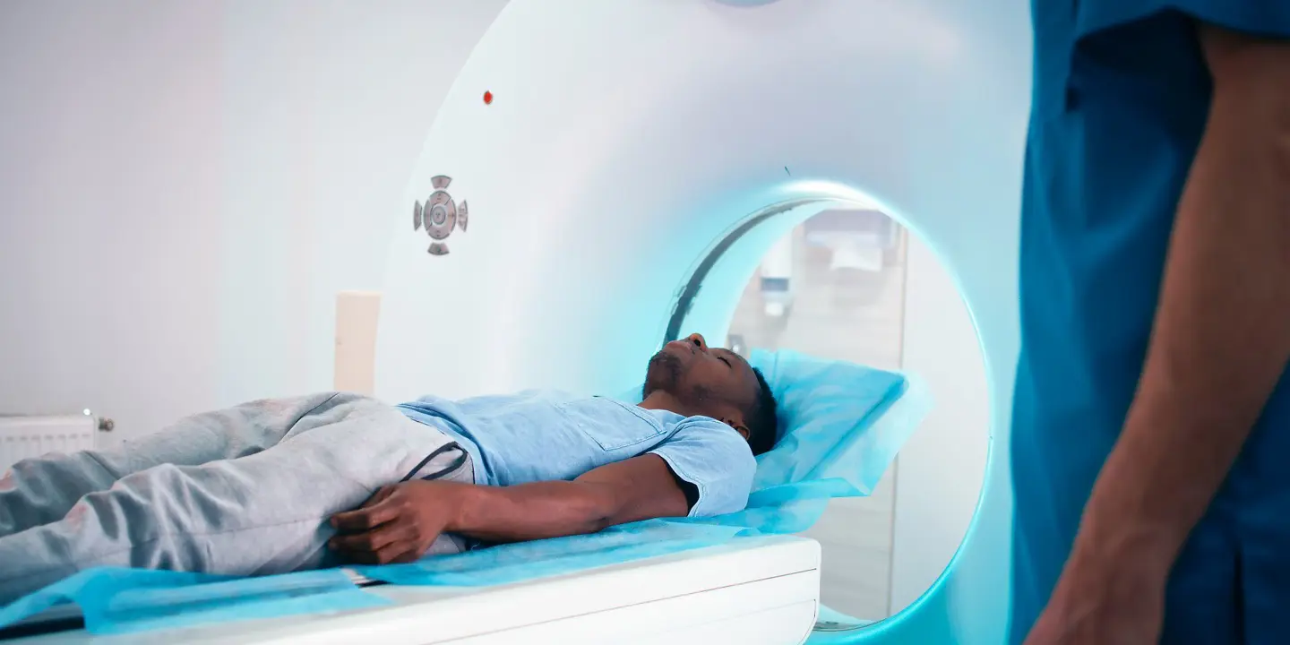
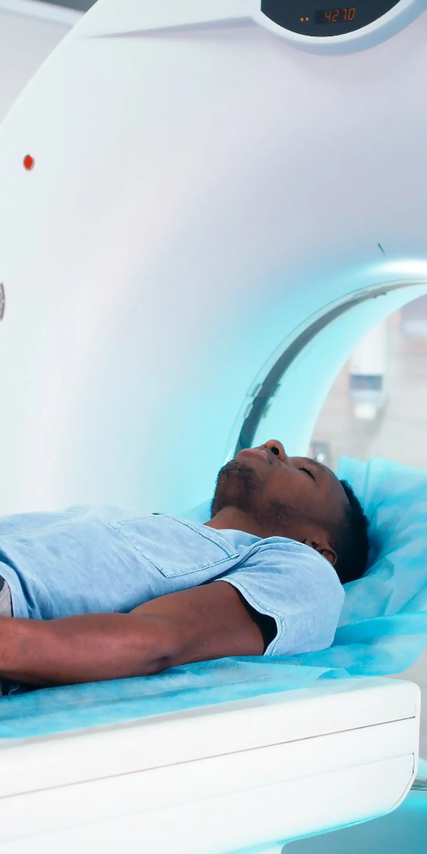
When you have a CT, a healthcare professional called a radiographer will ask you to lie back on a patient table or flat bed. The table will be slid into the CT scanner to perform your scan.
The CT scanner consists of a doughnut-shaped structure and a computer. The detectors in the scanner move around the part of your body being checked.
During the scan, you will lie on a comfortable bed that moves slowly through the doughnut-shaped part of the scanner. The process is painless and usually takes just a few minutes.
The doughnut structure contains an X-ray generator and an X-ray detector. As it rotates around your body, it releases a beam of painless X-rays that passes into your body.
The scanner detects how the X-rays move through your body to create clear pictures. This information is passed to a computer to create detailed images that form your scan results.
Different tissues absorb different amounts of X rays. Denser tissues absorb more and appear brighter on a CT image. Less dense tissues appear varying shades of grey, and the least dense areas (eg those filled with air, such as the lungs) appear black.
CT with contrast material
In some cases, your doctor may recommend a CT with a special dye called contrast.
Sometimes a harmless dye, called contrast, is used to make certain areas show up more clearly on the scan.How contrast dye is given depends on the part of your body that needs to be imaged.
Computed tomography scans mostly use iodine-based contrast materials. Iodine absorbs higher amounts of X-rays, so body parts flooded with iodine appear brighter and whiter on a CT scan (eg blood vessels, intestines, the stomach and fluid around the spinal cord) and reveal more detail.
Types of CT scan
There are several types of CT scans that are specific to the area of the body or internal organs that need to be investigated, such as abdominal, chest, joint and pelvic CT scans. Other types of CT include:
-
CT coronary angiogram – to visualise the blood supply to the heart (coronary arteries) and help detect heart disease
-
CT calcium score – to measure the amount of plaques in the coronary arteries
-
CT venogram – to visualise the veins, most often the veins in the pelvis and legs
-
CT myelogram – to visualise the spine; not only the bones but the spinal canal, spinal cord and nerves
Whichever type of CT scan you have, you will likely be asked to change into a hospital gown and should avoid wearing jewellery or other metal items on your body as these will interfere with the scan.
Determining the frequency of CT scans
If you have unexplained symptoms, such as pain, discomfort or swelling, a CT scan can help uncover the cause of your symptoms. CT scans can also be used to monitor the progression of a disease and track the effectiveness of a treatment (eg cancer treatment CT scans).
Factors influencing CT scan frequency include:
-
Whether your symptoms have changed – this may be in response to a treatment or over time without treatment
-
Your risk level for disease progression or complications
Because CT scans use X-rays, doctors are careful to recommend them only when needed. This avoids unnecessary testing and exposure to radiation. Your doctor will always weigh the small risks of radiation against the clear benefits of diagnosing or tracking a condition.
Health conditions requiring regular CT scans
A limited number of health conditions require regular CTs. How often depends on the specific health condition and your particular circumstances.
Health conditions that may require more than one CT to check for disease progression and/or the effectiveness of treatment include:
Cancer
Many different types of cancer are monitored before, during and after treatment using CT technology. This includes bowel, bladder, breast, lung, ovarian, prostate and womb cancer.
This helps determine how well cancer treatment, such as radiation therapy or chemotherapy, is working and provides evidence for whether different treatment options need to be explored.
Crohn’s disease
Crohn’s disease is a type of chronic inflammatory bowel syndrome that can affect any part of the gut. It usually goes through periods of high disease activity (flare-up) and low disease activity (remission).
You may need a CT if you experience a flare-up. This helps determine the extent of the flare-up and whether any complications have developed, such as a fistula, abscess, intestinal obstruction or ulcer.
Interstitial lung disease
This refers to over 200 different conditions that cause inflammation, thickening and scarring of the supportive tissue of the lungs. Regular scans monitor disease progression to get a clearer picture of the prognosis over time.
Risks and safety of frequent CT scans
CT scans use more ionising radiation than standard X-rays. This may have you wondering “are CT scans dangerous?” Broadly speaking, the answer is no.
The radiation level is low - roughly the same as you would naturally get from your surroundings over a couple of years.
Even so, regular scans are only recommended when there’s a medical need so you’re not exposed to extra radiation unnecessarily.
As well as radiation exposure, CT scans that use a contrast agent come with a small risk of developing an allergic reaction to the contrast material.
If you have been given a contrast agent before and have not developed an allergic reaction, you’re unlikely to develop an allergic reaction during future scans with contrast.
However, you will be monitored at the scanning facility for 30 minutes after your scan as most allergic reactions develop within minutes of having a contrast agent.
Comparing CT scans to other imaging tests
CT vs MRI
Both CT scanners and MRI (magnetic resonance imaging) produce detailed images of the inside of your body. However, MRI offers better contrast resolution for detecting subtle differences within soft tissues, while CT offers better spatial resolution.
MRI does not use any ionising radiation and instead uses powerful magnets and radio waves. This means MRI is generally safe to use during pregnancy, but it may not be suitable if you have a metal implant that is not compatible with MRI machines. MRI also takes longer to perform than a CT.
CT vs ultrasound
Unlike a CT, ultrasound does not use any ionising radiation and instead uses high-frequency sound waves. This allows ultrasound to create real-time images of the inside of your body whereas CT scan images are static. Ultrasound is also completely safe to use in pregnant women.
Ultrasound can’t provide detailed images of structures deep inside the body because of the limitations of sound waves when they encounter air or bone. CT provides highly detailed pictures with excellent spatial resolution, which means they can clearly define boundaries between different tissues.
CT vs X-ray
A CT and X-ray both use ionising radiation in the form of X-rays. An X-ray uses lower doses and is faster, but produces lower resolution 2D images. CT scans create cross sectional images through the area imaged and process them to form a highly detailed 3D image.
Do you need a CT scan?
If you have unexplained symptoms, it’s important to discuss these with a doctor so they can determine if an imaging test is needed. They can then advise you on whether a CT scan is the most suitable option.
At Vista Health, you can book a consultation with one of our experienced GPs who will discuss your symptoms and concerns. If appropriate, they can refer you for a CT scan at any one of our nationwide clinics.
Discover more about our advanced CT scans today.
Sources
https://www.radiologyinfo.org/en/info/safety-hiw_08
https://www.medicalnewstoday.com/articles/crohns-disease-ct-scan-vs-normal#uses
https://pmc.ncbi.nlm.nih.gov/articles/PMC9537704/
https://publications.ersnet.org/content/errev/34/176/240194
https://pmc.ncbi.nlm.nih.gov/articles/PMC4164599/
https://my.clevelandclinic.org/health/diseases/17809-interstitial-lung-disease
https://www.radiologyinfo.org/en/info/safety-contrast
https://www.insideradiology.com.au/iodine-containing-contrast-medium/
https://www.cancerresearchuk.org/about-cancer/tests-and-scans/ct-scan
https://www.bhf.org.uk/informationsupport/heart-matters-magazine/medical/tests/ct-scans-of-the-heart
https://my.clevelandclinic.org/health/diagnostics/24929-venogram


