
Clinically reviewed by Peter Kabogoza
National Clinical Lead - Ultrasound
Thyroid Ultrasound: Grading Nodules (U1 to U5)
Around 4 in 10 adults in the UK have thyroid lumps, with most occurring in women. These lumps, also known as thyroid nodules, are usually not cancerous. However, in around 1 in 20 cases, a thyroid nodule is thyroid cancer.
If you feel a lump in your neck, a thyroid ultrasound scan is an important step to investigate your risk of thyroid cancer.
Here, we’ll explore how thyroid nodules are categorised, what a private thyroid ultrasound scan can show and when you might need one.
Understanding thyroid nodules and ultrasound grading
What is the thyroid?
Your thyroid gland is a butterfly-shaped gland in the front of your neck. It makes thyroid hormones, which are essential for a variety of bodily functions. This includes your metabolism, energy levels, heart rate, body temperature and calcium levels.
What are thyroid nodules?
Thyroid nodules are abnormal growths of tissue in the thyroid gland. These lumps are commonly benign (not cancerous) and can range from one to several centimetres in diameter.
They are not the same as goitre. Goitre is when the entire thyroid gland is enlarged. Goitre sometimes involves a few or many nodules, but it can also present without any nodules.
Why is ultrasound important for neck lump scan?
An ultrasound examination is a quick, non invasive, painless scan for investigating neck lumps. It does not involve the use of special dyes (contrast agents) or radiation.
It can identify the boundaries between tissues of different densities, allowing your doctor to determine whether your neck lump is solid, fluid filled (a cyst) or a mixture. Solid lumps are the most likely to be cancerous, although most solid lumps are usually not thyroid cancer.
Thyroid nodule classification systems
When you have ultrasound imaging of a thyroid lump, your ultrasound practitioner will categorise the lump according to an established grading system.
In the UK, the most commonly used system is the British Thyroid Association system, namely BTA U. In the USA, the TI-RADS system is more commonly used.
The TI-RADS system is a points-based system where 5 different features of the nodules are assessed, and each receives a certain number of points. The points are added together to give you a final TI-RADS score.
Both TI-RADS and BTA U are effective systems to diagnose thyroid problems.
BTA U classification system overview
The BTA system has 5 grades. The first grade, U1, is normal, where no nodules are detected in the thyroid.
The next 4 grades all refer to the detection of nodules. The higher the grade, the more likely the nodule is cancerous based on the features picked up during the scan. The grades are as follows:
-
U2 – benign; features suggest it’s not cancerous
-
U3 – indeterminate; can’t determine based on features alone whether it’s likely to be cancerous or not
-
U4 – suspicious; features suggest there’s a high risk that it’s cancerous
-
U5 – malignant; features suggest there is a very high risk that it’s cancerous
U3 to U5 all refer to suspected thyroid disease, which will need further diagnosis.
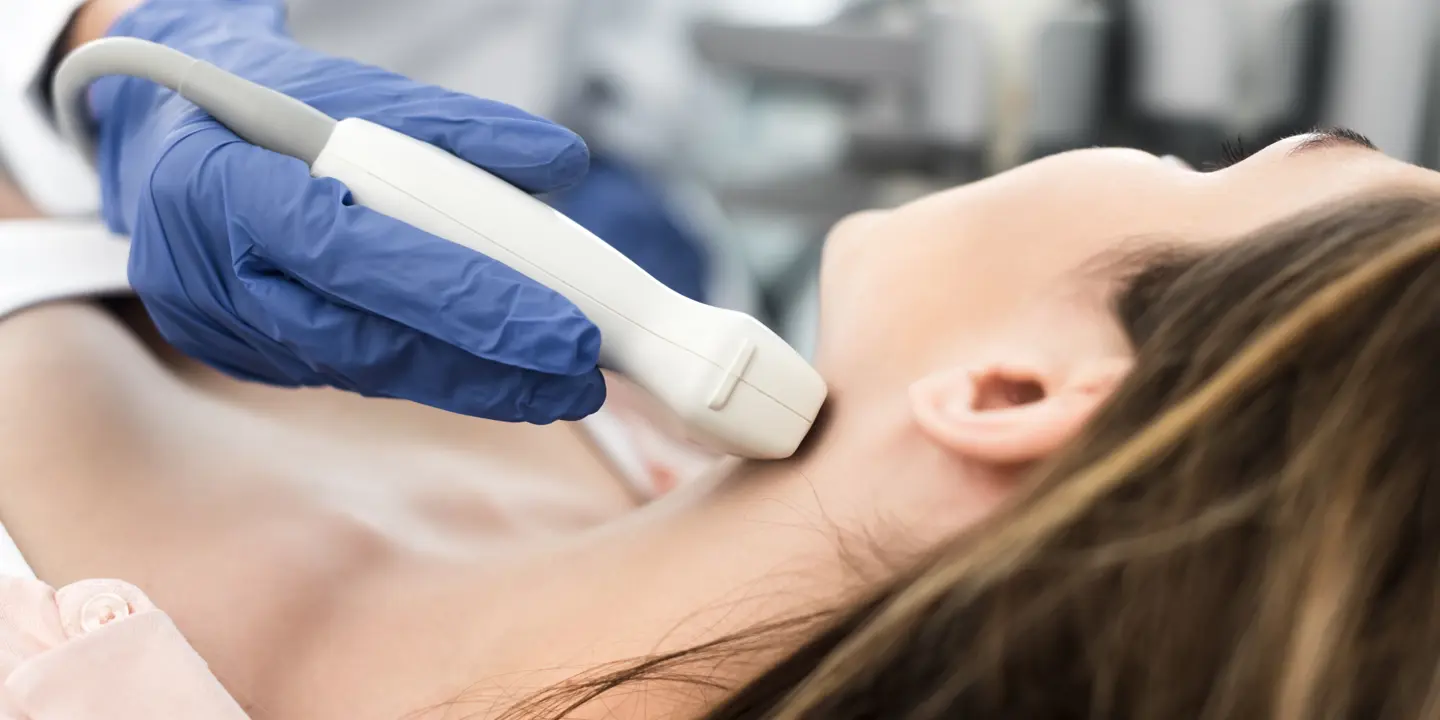
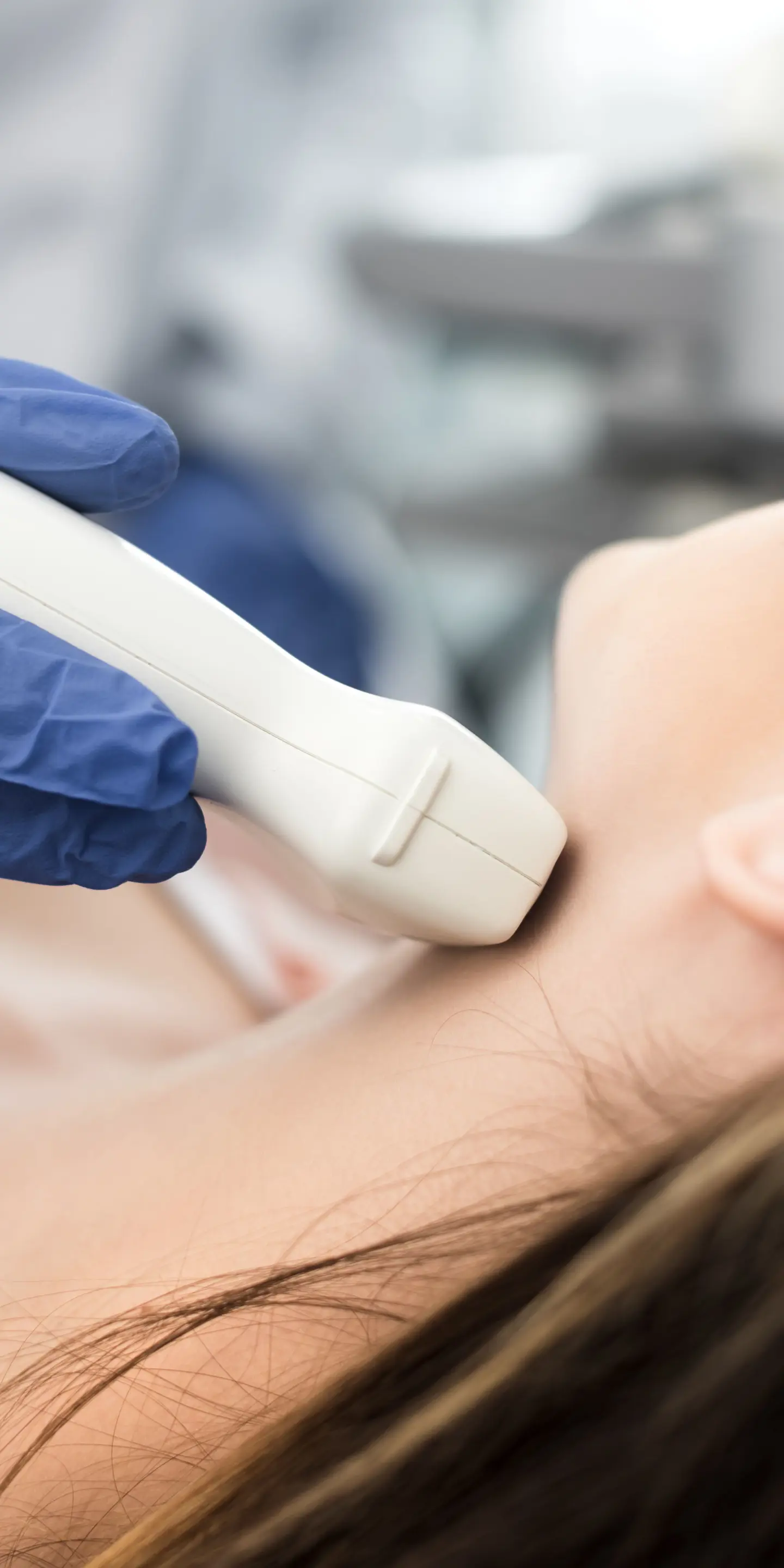
How thyroid ultrasound works in nodule evaluation
An ultrasound thyroid scan uses high frequency sound waves to create images of soft tissues in the body. The ultrasound machine is a small, portable device with a monitor and a handheld ultrasound probe.
During a thyroid scan, ultrasound waves are released by the handheld probe into the neck. The probe is moved around the neck to assess the thyroid from different angles. The sound waves bounce back whenever they encounter a boundary between tissue areas of different densities.
The bounced sound waves are detected by the ultrasound probe. Over 1,000 of these bounced waves can be detected in one second. This allows ultrasound to produce live, 2D images of the inside of the body.
A thyroid ultrasound scan typically takes between 15-20 minutes.
When is a thyroid ultrasound recommended?
A thyroid ultrasound scan is recommended if you have symptoms such as swelling of the neck, a lump in your neck, or, in some cases, if you have an abnormal thyroid function test.
A doctor may also recommend that patients have a thyroid ultrasound scan based on a combination of their medical history and symptoms associated with thyroid disorders. This includes unintentional weight changes, poor control of body temperature, extreme tiredness, poor sleep, and unexplained hair loss and/or dry skin.
What happens after your thyroid ultrasound
You can return to your usual activities immediately after your scan. At Vista Health, we aim to email you a report with your results within 3 working days. It can also be sent to your NHS GP.
Your report will be overseen by one of our experienced Consultant Radiologists. You can then follow up with your NHS GP or book a private GP consultation with a Vista Health GP. They will explain what your results mean for you and any next steps.
If your thyroid scan suggests you may have thyroid problems and/or your lump is graded as U3 or above, you will get a GP referral to a specialist.
Your specialist will likely recommend other tests, such as blood tests to assess your thyroid function, and in some cases, fine needle aspiration. This is a type of biopsy, where a very thin needle is inserted into the nodule to collect cells for analysis under a microscope. A biopsy is the only way to reach a diagnosis of thyroid cancer with complete certainty.
Depending on your results, your doctor will recommend appropriate treatment.
Get clarity on your thyroid health
If you’re worried about swelling or a lump in your neck, find the answers you are looking for with a private ultrasound scan of your thyroid at one of Vista Health’s nationwide clinics.
You’ll need a GP referral for this scan, which you can obtain either from your NHS GP or through a private virtual GP consultation with us.
Sources
https://pmc.ncbi.nlm.nih.gov/articles/PMC6987506/
https://my.clevelandclinic.org/health/diagnostics/thyroid-ultrasound
https://radiopaedia.org/articles/bta-ultrasound-u-classification-of-thyroid-nodules