
Clinically reviewed by Peter Kabogoza
National Clinical Lead - Ultrasound
Ultrasound Scan: What It Can Detect in Your Body
Ultrasound scans use sound waves to create images of the inside of your body.
Ultrasound images are most often used to detect changes in soft tissues, such as muscles, ligaments and tendons, internal organs, blood vessels, lymph nodes, and glands such as your thyroid gland. They also detect abnormal growths such as fluid-filled sacs or cysts. They’re used alongside your medical history to investigate symptoms and diagnose conditions causing unexplained pain or discomfort.
Here, we will explore how ultrasound scans work and common conditions they can detect.
How ultrasound scans detect issues
Ultrasound, also called sonography, uses high frequency sound waves that are beyond the range of human hearing.
The sound waves are released by a small device called an ultrasound transducer (or ultrasound probe). When they pass into your body, the sound waves bounce back when they encounter a body issue.
The reflected sound waves are detected by the handheld transducer as it passes over your body. This information is transmitted to a computer as electrical signals, which vary depending on the stiffness and density of the tissue they’re bouncing off. The electrical signals are then turned into greyscale images.
In just one second, over 1,000 sound waves can be reflected, meaning images of the inside of your body are created in real-time and displayed on a computer screen as your scan is carried out.
Two advantages of ultrasound over other tests and imaging techniques are that it doesn’t involve radiation and it can capture images while you move your body. For example, when investigating joint pain, your doctor can see how the tissues in your joint respond as you move your joint.
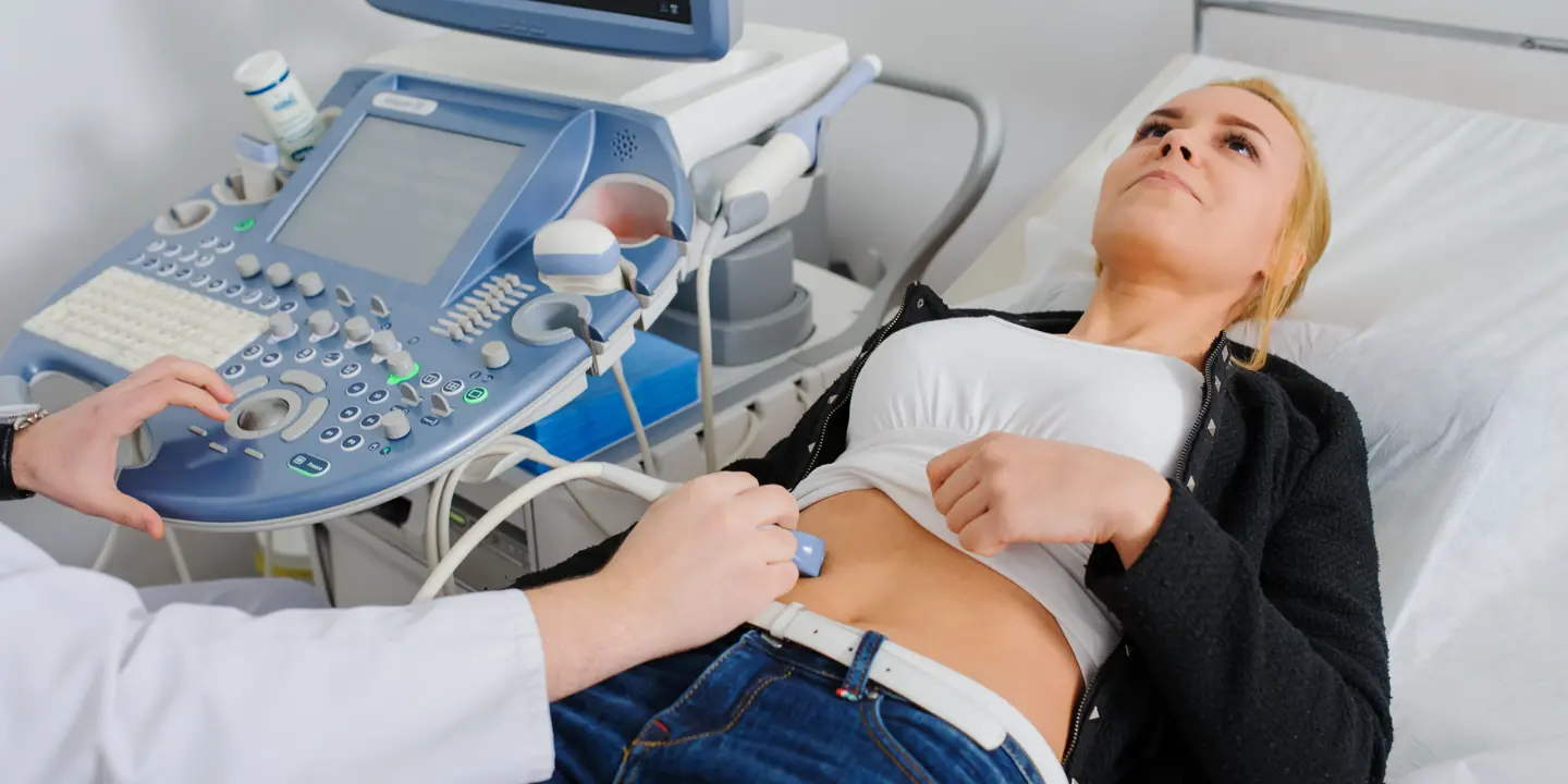
What to expect during an ultrasound scan?
Ultrasound machines are much smaller than machines used for CT scans or MRIs. They’re portable and use a handheld scanning device, which means scans can be performed while you’re sitting up, lying down or turned on one side of your body depending on which part of your body is being scanned.
Before your scan starts, you may be asked to remove your clothing and wear a hospital gown. Next, the skin over the part of your body being imaged will be cleaned.
A water-based gel will be applied to your skin where the imaging tests will take place. This allows for smooth contact between your body and the ultrasound transducer. It prevents air pockets that can interfere with the passage of sound waves from the transducer into your body and back again.
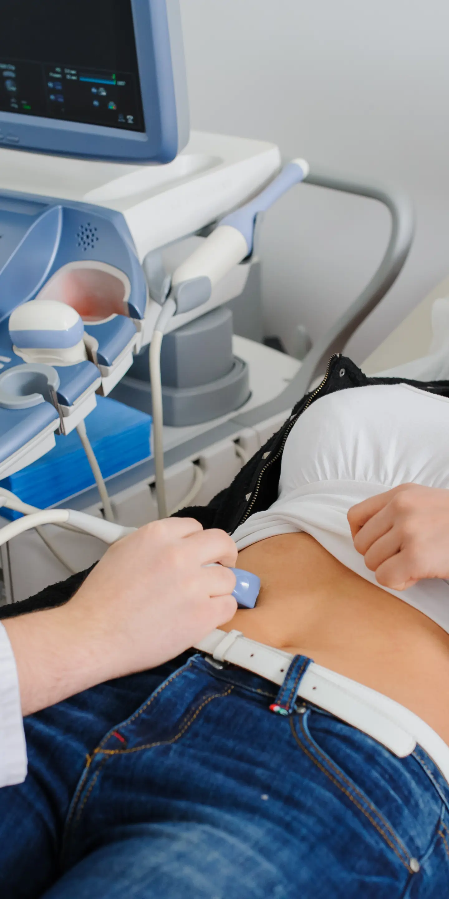
A specially trained clinician called a sonographer or ultrasound practitioner will move the transducer along the part of your body to be imaged. They may need to apply a small amount of pressure to obtain clearer images. This does not usually hurt but may cause mild discomfort.
Your ultrasound practitioner may also tilt the transducer to better capture images of your tissues from different angles for a more complete view. For most ultrasound scans, you will be able to see the images created in real-time on a computer screen.
Your ultrasound scan can take from 15 to 45 minutes, depending on which part of your body needs to be imaged. Once your ultrasound scan is complete, you can return to your usual activities straight away. At Vista Health, we aim to deliver your scan results within 3 working days.
Common conditions identified by ultrasound
Ultrasound scans are useful in diagnosing a wide range of conditions because they can:
-
assess organ health
-
detect abdominal or pelvic problems
-
identify vascular and blood flow issues
What’s more, ultrasound scans are safe to use for most people as they have no known risks and do not:
-
involve being passed into a scanning machine – this can be challenging for individuals with claustrophobia or anxiety
-
use ionising radiation, making them safe for children and pregnant women
-
use strong magnets, making them safe for those with metal implants
Diagnostic ultrasounds are often used to help detect disease and damage early, which allows for fast treatment.
What can an ultrasound scan of the abdomen detect?
Ultrasound scans of the abdomen are commonly used to investigate abdominal pain that may be related to your liver, kidneys, pancreas, gallbladder or urinary bladder. This is different from a bowel ultrasound, which identifies problems with the digestive system, including the stomach and intestines.
An abdominal ultrasound scan can be used to detect growths that may need to be tested for liver cancer, for example. It can also help diagnose other liver diseases such as hepatitis, fatty liver disease and liver scarring or cirrhosis.
When used to investigate the kidney, an abdominal ultrasound can help detect:
-
kidney masses that may be suspicious of cancer
-
kidney cysts – fluid-filled sacs
-
kidney stones – hard stones made of minerals and salts
-
kidney infections (pyelonephritis) – for example, infection caused by the bacterium E.coli travelling up into the kidneys from the bladder or urinary tract
An abdominal ultrasound can also help detect inflammation of the pancreas (pancreatitis) and possible pancreatic cancer.
When used to investigate the gallbladder, it can help detect:
-
gallstones – hard stones made of bile pigments, calcium salts and cholesterol
-
blockage of the bile duct
-
inflammation of the gallbladder (cholecystitis)
Although the urinary bladder sits in the pelvis, when full, it expands into the abdomen. As it is easier to image the bladder clearly when it’s full, you’ll usually be asked to drink several glasses of water before an ultrasound scan of your bladder.
A bladder ultrasound can help diagnose:
-
an overactive bladder – a condition that causes sudden urges to urinate and interferes with your quality of life
-
bladder cancer
-
bladder stones – hard stones made of minerals and salts
What can an ultrasound of the pelvis detect?
Pelvic ultrasound scans are used to investigate the reproductive organs of men and women.
The ultrasound device used for these investigations may differ from a standard transducer.
For example, in women, a pelvic ultrasound may involve the use of a transvaginal ultrasound transducer that is inserted into the vagina. Similarly, in men, a transrectal transducer may be used. This is inserted into the rectum to investigate the prostate. These are examples of internal ultrasound scans, which are carried out with probes designed for insertion, compared to external ultrasound scans, where the transducer is passed over your skin.
In women, an ultrasound scan of the pelvic organs can help detect:
-
an abnormally shaped womb
-
ectopic pregnancy
-
uterine fibroids – non-cancerous growths in the uterus (womb) made of fibrous tissue and muscle
-
ovarian cysts (fluid-filled sacs)
-
pelvic inflammatory disease (PID) – infection and inflammation of the female genital tract (womb, ovaries and fallopian tubes)
-
uterine cancer
A pelvic ultrasound scan in women, also known as a gynaecological ultrasound, can help diagnose polycystic ovarian syndrome (PCOS). This condition causes a large number of fluid-filled sacs called follicles on the ovaries.
In men, an ultrasound scan of the pelvis can help detect:
-
an enlarged prostate, also known as benign prostatic hyperplasia (BPH)
-
inflammation of the prostate (prostatitis)
-
prostate and testicular cancer
-
testicular cysts (fluid-filled sacs)
-
testicular torsion – twisting of the cord supplying blood to the testicles
-
varicoceles – enlarged veins of the sac that contains the testicles (scrotum)
What can an ultrasound of the heart detect?
An ultrasound of the heart is called a cardiovascular ultrasound, doppler ultrasound or echocardiogram. It investigates the structure and function of the heart and the main blood vessels that supply it. It can also evaluate blood flow through the heart.
A cardiovascular ultrasound can help detect:
-
cardiomyopathy – thickening of the muscular wall of the heart
-
endocarditis – infection of the inner lining of the heart
-
heart attack damage
-
heart defects
-
heart failure
-
heart valve problems
What can an ultrasound of the joints and surrounding soft tissues detect?
As ultrasound scans allow for real-time imaging of tissues, they are often used to investigate joint and musculoskeletal problems to determine how movement affects these tissues.
An ultrasound scan of the musculoskeletal system can help detect:
-
bursitis – inflammation of the fluid-filled sacs that enable smooth movement of the joints (bursae)
-
ganglion cysts – fluid-filled sacs near joints
-
ligament injuries – this includes tears of the collateral ligaments in the knee and sprains of the lateral ligament complex in the ankle
-
muscle injuries – this includes muscle growth due to damage, fluid build-up (oedema) and tears
-
tendonitis – inflammation of the tendons, including iliotibial band syndrome, where the tendon running from the hip to the knee becomes inflamed
-
tendon tears – this includes tears of the:
-
achilles tendon in the ankle
-
bicep tendon in the upper arm
-
patellar tendon in the knee
-
rotator cuff in the shoulder joint
-
When to consider getting an ultrasound scan
Ultrasound exams are a quick, painless and safe way to investigate symptoms, such as discomfort, unexplained pain, swelling or tenderness.
If you have any of these symptoms and do not want to wait to have them investigated, you can self-refer for an ultrasound scan with Vista Health. You do not need to obtain a referral from a healthcare professional.
Ready to get clarity on your health? Book your private ultrasound at one of Vista Health’s nationwide clinics.
Sources
https://www.mountsinai.org/health-library/report/gallstones-and-gallbladder-disease
https://my.clevelandclinic.org/health/diagnostics/4994-abdominal-ultrasound
https://www.nhs.uk/conditions/endometriosis/
https://www.hopkinsmedicine.org/health/treatment-tests-and-therapies/pelvic-ultrasound
https://www.radiologyinfo.org/en/info/us-prostate
https://www.radiologyinfo.org/en/info/us-scrotal
https://my.clevelandclinic.org/health/diagnostics/13477-echocardiogram-transthoracic-tte
https://my.clevelandclinic.org/health/diagnostics/16947-echocardiogram