
Clinically reviewed by Raphael Owononi
Radiology and Radiation Protection Clinical Lead

When to Get a Chest X-Ray for a Persistent Cough
If you’re wondering “should I get a chest X-ray for a cough?”, the answer partly depends on how long you’ve had symptoms for. In general, if you’ve had a persistent or chronic cough (3 weeks or longer), you should see a GP to determine whether a chest X-ray is needed.
Here, we’ll take a look at when to get a chest X-ray for a cough, how a chest X-ray can help diagnose lung problems, and the risks and benefits of having a chest X-ray.
We will also explore different types of coughs and their causes, and other symptoms, as well as other treatments for diagnosing lung conditions. First, it helps to understand how an X-ray works.
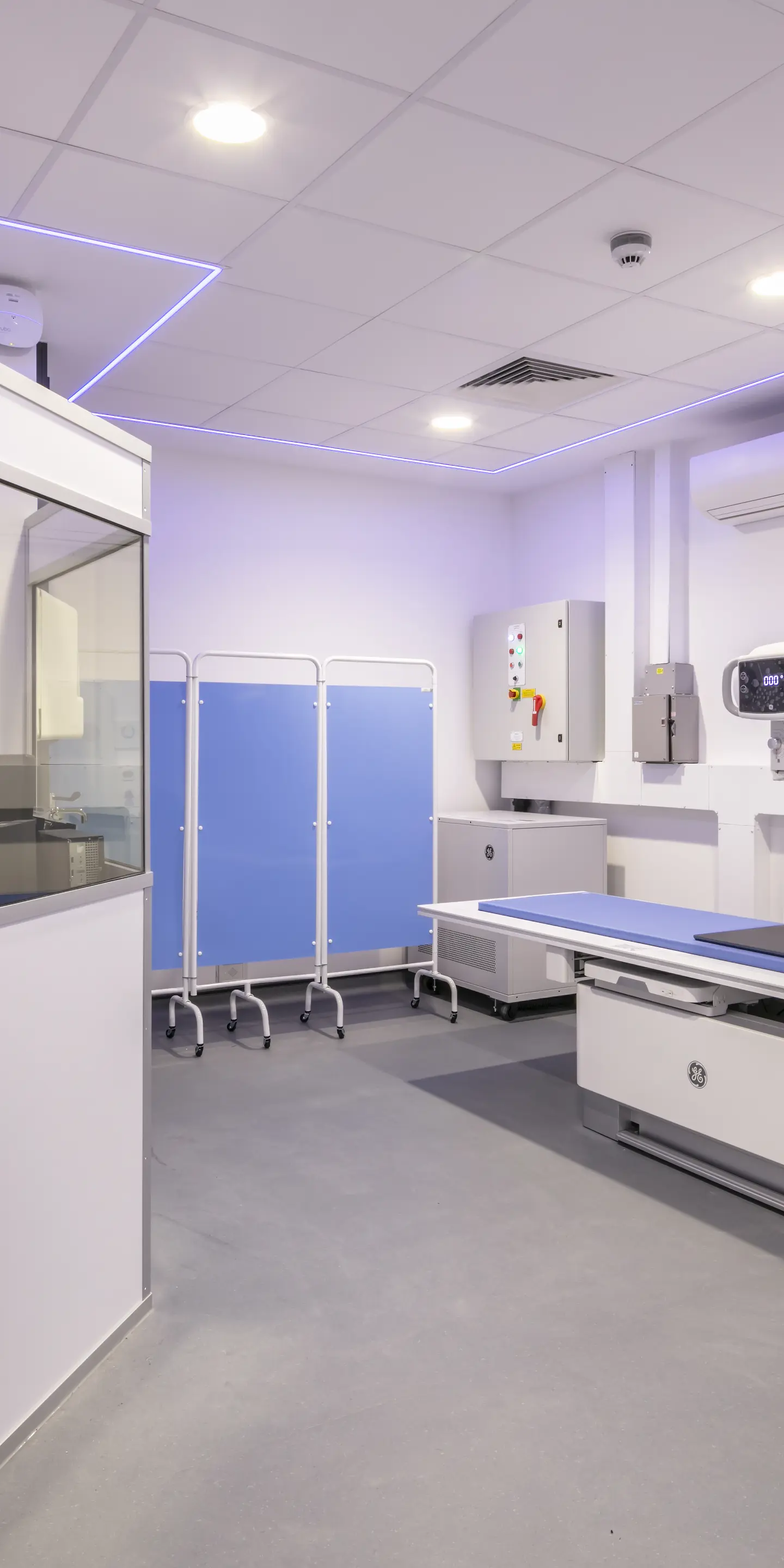
What is a chest X-ray?
A chestX-ray is a type of medical imaging that is safe, painless and non-invasive. It uses low doses of ionising radiation to create images of the inside of your chest cavity.
A chest X-ray, or chest radiograph, focuses on your lungs, heart, ribs and other tissues in your chest. A single chest X-ray uses 0.1 mSv of radiation, which is equivalent to 10 days of exposure to the radiation present in our natural environment.
An X-ray machine works by sending beams of X-ray radiation through your body. Different tissues in your body absorb different amounts of X-rays. The X-ray radiation that passes through your body and is not absorbed is picked up by an X-ray detector.
The X-ray detector converts the X-ray signal into an electrical signal that is sent to a computer. This is used to create a digital image.
Tissues that are dense (eg your bones) absorb more X-rays and so appear whiter on an X-ray image. Tissues that absorb fewer X-rays (eg your fat, muscles and organs) appear in varying shades of grey. Air appears black on an X-ray image.
Signs you need a chest X-ray for a cough
So, when should you get a chest X-ray for a cough? The short answer is when you have a persistent or chronic cough. This means a cough that lasts for 3 weeks or more. It could be a dry cough, hacking cough or a wet, chesty cough.
As most coughs get better on their own within 3 weeks, any lingering cough should be investigated as soon as possible. This is particularly important if your persistent cough occurs alongside:
-
chest pain when breathing or coughing
-
coughing up blood (haemoptysis)
-
dizziness and/or lightheadedness
-
heart palpitations ie feeling that your heart is pounding or racing
-
nausea and/or vomiting
-
difficulty breathing, such as shortness of breath
Other signs that you should urgently consult your doctor about a persistent cough include:
-
peeing less than is normal for you
-
a very high temperature
-
a very low temperature – this can be a sign that your immune system is unable to fight off a chest infection (eg pneumonia)
Understanding cough types and their causes
A cough is your body’s natural response to things that are irritating your airways – this includes your throat and the tubes that branch out into your lungs (bronchi). Coughing is your body’s attempt to remove irritants from these places.
A persistent or chronic cough can, therefore, be a sign that there is an ongoing problem affecting your airways. Coughs can be categorised according to how long they last, the symptoms they produce and how they sound.
Acute vs persistent vs chronic
Acute coughs last less than 3 weeks, while persistent coughs last more than 3 weeks. Chronic coughs last for 8 or more weeks.
Wet vs dry
A productive or wet cough brings up phlegm (thick mucus produced by your throat and lungs) while a non-productive or dry cough does not. A wet cough is usually the result of a lung infection, which can be caused by:
-
a common cold
-
bronchitis – one cause of bronchitis is post-nasal drip, which occurs due to acute sinusitis
-
influenza
-
pneumonia
-
tuberculosis
You may experience a dry cough at the start of a lung infection before the airways are very irritated and at the end of a lung infection when the airways are recovering. This is often the case with acute bronchitis.
A persistent dry cough without any associated wet cough can be caused by asthma, allergies or acid reflux caused by gastro-oesophageal reflux disease (GORD).
Wheezing vs whooping
A cough that sounds like a wheeze is usually due to the airways being partially blocked. This can occur with an infection or asthma, both of which cause the airways to swell and produce excess mucus.
A cough that sounds like a whoop is caused by pertussis, which is also known as whooping cough. This is caused by infection with the bacteria Bordetella pertussis.

How chest X-rays help diagnose respiratory issues
A chest X-ray with no significant findings will show healthy lungs that appear clear.
In contrast, a chest X-ray with significant findings will reveal lungs that are damaged, diseased or infected will have abnormalities that appear as bright masses, white spots or hazy patches in the chest cavity.
These can be signs of fluid or pus build-up, growths or scar tissue. A chest X-ray can, therefore, help detect:
-
abscesses – pus-filled cavities that often develop due to an infection
-
cysts – air-filled sacs that can develop with age but can also be a sign of disease
-
infections
-
nodules (masses 3cm or less in size) and tumours (masses greater than 3cm in size)
-
pleural effusions – fluid collection around the lungs specifically in the space between 2 thin layers that surround the lungs called the pleural membranes
-
pulmonary fibrosis – scarring of the tissue that surrounds the air sacs of the lungs
A chest X-ray can also detect broken ribs and problems with your heart, such as:
-
an enlarged heart
-
fluid build-up in your heart that may be a sign of heart failure
-
calcium deposits in your heart and the blood vessels that supply it, which may suggest heart disease
Comparing chest X-rays with other diagnostic methods
A chest X-ray is a quick, cost-effective and non-invasive tool for detecting lung problems.
They are frequently used after a chest injury to identify broken bones and/or a collapsed lung. However, they are also often used to rule out conditions such as pneumonia or heart failure. This can help your doctor more easily identify the cause of your symptoms.
Consequently, after having a chest X-ray, your doctor may recommend further diagnostic tests, such as lung function tests, a lung CT scan and/or collection of a lung tissue sample (biopsy).
A lung CT scan provides greater detail than a chest X-ray and can, therefore, be used to accurately diagnose lung conditions such as chronic obstructive pulmonary disease (COPD) and emphysema. Lung CT scans can also better assess damage caused by cystic fibrosis.
As a lung CT scan can detect subtle differences within tissues, it can provide a clearer picture of the severity and type of lung condition.
Due to its higher resolution, a lung CT scan can also help provide a definitive treatment diagnosis when it comes to masses in the lungs. For example, a normal chest X-ray can detect a suspicious mass but may not be able to determine if it’s lung cancer or a lung abscess. However, a lung CT scan can provide a clear diagnosis.
Potential risks and benefits of chest X-rays
Chest X-rays are a low-risk, medical imaging test. They are quick, taking only 5–10 minutes, while a lung CT scan can take up to 30 minutes.
A chest X-ray exposes you to a very low dose of radiation – equivalent to 10 days of exposure to background radiation in the environment. In contrast, the amount of radiation that a lung CT scan exposes you to is the equivalent of 2 years of background radiation (around 6.1 mSv).
However, a chest X-ray only provides 2D images of the inside of your chest and these images do not offer clear details of soft tissues. Consequently, depending on the findings of your chest X-ray, you may need further imaging services such as a lung CT scan and other treatments.
What to do after your chest X-ray
At Vista Health, we aim to provide your chest X-ray results within 3 working days. This will include a detailed report from one of our experienced consultant radiologists. You can then discuss your results with one of our expert GPs or alternatively, we can send the results to your NHS GP.
When you choose to consult a Vista Health GP, you can rest assured that your results will be clearly explained, with advice specific to your particular circumstances. We will advise you on your options so you can make an informed decision on next steps to get your lung health back on track with the right treatment.
Do you need a chest X-ray?
If you’re concerned about a cough that won’t go away, book a consultation with one of our dedicated private GPs.
Based on your symptoms, concerns and medical history, they will let you know whether a chest X-ray could help. They can then help arrange a private X-ray with Vista Health.
Learn more about our private X-ray service today.
Sources
https://www.radiologyinfo.org/en/info/safety-xray
https://www.nhsinform.scot/illnesses-and-conditions/lungs-and-airways/pneumonia/
https://my.clevelandclinic.org/health/diagnostics/10228-chest-x-ray
https://www.nhsinform.scot/illnesses-and-conditions/lungs-and-airways/cough/
https://www.mayoclinic.org/tests-procedures/chest-x-rays/about/pac-20393494
https://my.clevelandclinic.org/health/symptoms/17755-cough