 Clinically reviewed by Liz Marsh
MRI Clinical Lead
Why Do I Need an MRI Scan for Gallstones?
Gallstones are small, hard, lumps that form in the gallbladder. While they don’t usually cause symptoms, in some cases, gallstones can cause severe pain in your abdomen. Pain in your tummy can be caused by many different things, so it’s important to accurately diagnose what’s causing your pain.
A gallbladder MRI scan can help diagnose the cause of your pain, including if you have gallstones. If gallstones are ruled out as the cause of your unexplained abdominal pain, an MRI scan can help you get to the bottom of your symptoms sooner.
Read on to learn what gallstones are and their symptoms. We’ll also explore how MRI detects gallstones, other ways of having gallstones diagnosed, and how these compare to MRI.
Understanding gallstones and their symptoms
Gallstones are small stones your body makes from substances found in bile – a thick, yellow-green fluid that is made by your liver and stored in your gallbladder. Bile contains:
-
Cholesterol
-
Bile salts – also known as bile acids
-
Bilirubin – a red-orange breakdown product of the pigment heme found in red blood cells
-
Body salts – such as calcium, sodium, potassium and magnesium
-
Metals such as copper
-
Phospholipids – a type of fat
Your gallbladder is part of your digestive system. It sits underneath your liver and is connected to both your liver and small intestine through a network of tubes called bile ducts. These ducts eventually merge together to form the common bile duct, which transports bile into the small intestine.
Bile has two main roles in your body:
-
To help digest and absorb fat from your food
-
To carry waste products made by your liver out of your body via your stools
If there is an imbalance in the substances that make up your bile, hard deposits start to form, i.e. gallstones. Most people will not develop any symptoms of gallstones – these are called silent gallstones. However, if a gallstone blocks one of your bile ducts, it can interrupt bile flow through the duct.
If bile flow is disrupted, your gallbladder can become sore and tighten – this is called biliary colic. Biliary colic causes sudden, severe tummy pain and cramping that come and go.
Abdominal pain is usually located on the upper right side of the abdomen and can spread to your back and shoulder.
You may also experience:
-
Nausea
-
Sweating
-
Tenderness around your abdomen
-
Vomiting
How MRI scans detect gallstones
An MRI (magnetic resonance imaging) scan uses powerful magnets and radiofrequency waves to create highly detailed images of the inside of your body in 3D.
MRI scanners are made up of a patient table that slides into a tube. The tube contains powerful magnets as well as radiofrequency coils that produce radio waves.
The magnets create a strong magnetic field around you. This affects water molecules that are found in all the cells in your body, specifically tiny particles in water called protons.
The MRI scanner then sends gentle pulses of radio waves that affect the water molecules in your cells. This causes the water molecules to release signals that are detected by the MRI scanner. Areas of your body that hold more water show up brighter on the scan.
Magnetic resonance imaging is, therefore, able to scan all tissues in your body and produce detailed greyscale images. This allows MRI to detect tissue changes caused by disease or injury.
A special type of MRI scan is used to create clear and detailed images of the gallbladder and ducts, as well as the pancreas and liver. This is called magnetic resonance cholangiopancreatography (MRCP).
MRCP uses computer processing tailored specifically for imaging this area of the body. This makes it easier to detect gallstones and other conditions affecting the gallbladder.
When is an MRI scan recommended?
If you are experiencing symptoms of gallstones, your doctor may recommend blood tests to check for signs of inflammation and infection, as well as an abdominal ultrasound scan.
An ultrasound scan may be inconclusive i.e. it may not be clear from the abdominal ultrasound scan whether or not you have gallstones. An MRI scan may then be recommended to confirm or rule out gallstones or other digestive and kidney diseases.
Comparing MRI with other imaging techniques
Other gallbladder imaging tests that can help diagnose gallstones or investigate complications caused by gallstones include ultrasound and CT (computerised tomography).
MRI vs ultrasound
An abdominal ultrasound is often recommended first to investigate symptoms of gallstones as it is quick to perform and less expensive than an MRI. Ultrasound uses high frequency sound waves rather than strong magnets. This makes it suitable for individuals with metal implants that are not compatible with MRI machines.
However, due to the lower resolution of ultrasound compared to magnetic resonance imaging, it’s not always able to detect small gallstones. This lower resolution also means it may not be able to easily detect complications of gallstones or conditions other than gallstones that produce similar symptoms.
MRI vs CT
CT uses computer technology and ionising radiation in the form of X-rays to image the inside of your body. Although it provides higher resolution images than ultrasound, the nature of CT means it is less sensitive at detecting gallstones, so a CT scan is more commonly used to diagnose gallstone complications.
However, as CT uses X-rays it is not suitable for pregnant women. An Ultrasound or MRI scan is a safer imaging test for pregnant women.
Benefits of choosing an MRI scan
Comprehensive gallbladder imaging
An MRI scan is an imaging test that can simultaneously:
-
Detect small gallstones
-
Diagnose gallstone complications – complications include:
-
Acute pancreatitis – sudden inflammation of the pancreas
-
Cholecystitis – inflammation of the gallbladder
-
Jaundice – yellowing of your skin and the whites of your eyes due to a build-up of the pigment bilirubin in your blood
-
Diagnose other conditions that produce symptoms similar to gallstones e.g. gallbladder cancer (gallbladder carcinoma) and bile duct cancer
No ionising radiation
Unlike CT, an MRI exam does not use any ionising radiation, which makes it suitable for pregnant women.
High-resolution images
MRI images offer the highest contrast resolution of all medical imaging tests. This makes it ideal for detecting subtle changes within tissues e.g. for the detection of very small gallstones in the biliary tract, specifically the gallbladder and bile ducts.
What to expect during your MRI scan
Before your appointment, you will be asked to fast (not eat or drink) for 4 hours. During this time, you can have sips of water and any medication you would normally take. When you arrive for your MRI scan, you may be asked to change into a hospital gown. You will also be asked to take off any metal items on your body, such as metal piercings, metal jewellery and any metal medical devices that can be removed. This is because metal can distort areas of the images created or pose a hazard.
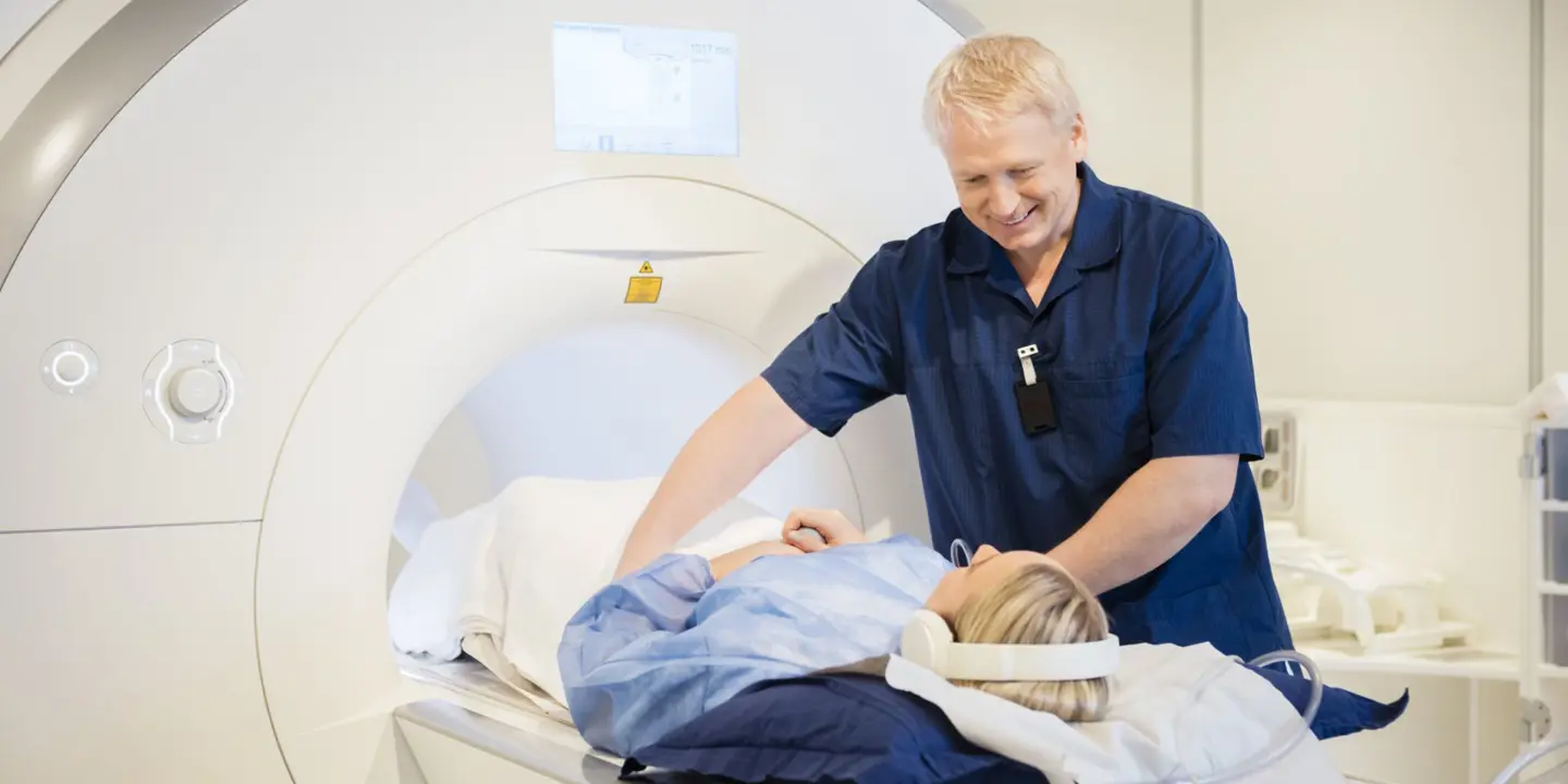
You will be asked to lie on your back on the patient table, with a pillow under your head, and will be slid into the MRI machine so your tummy is in the middle to begin your scan. Once you are in the MRI scanner, you will still be able to communicate with your care team via a buzzer and two-way intercom.
If you are feeling agitated or anxious for any reason, you can ask for the scan to be stopped. It helps to practice mindfulness techniques to help reduce your anxiety if you struggle with being in confined spaces. We can also give you headphones so you can listen to music during your scan.
You will need to remain still during your MRI to ensure clear images are created. Moving causes blurring on the pictures. During your scan, you will hear harmless loud banging, clicking and knocking sounds, which are caused by the normal processes of the MRI machine. We will give you earplugs to make them quieter.
After about 40 minutes, when your scan is complete, you can return to your usual activities straight away.
Interpreting MRI scan results for gallstones
At Vista Health, we aim to provide a detailed report of your MRI scan results within 3 working days. This report will be sent to you and can additionally be sent to your NHS GP. It will include insights from one of our experienced Consultant Radiologists.
This report can include findings such as whether or not you have gallstones, the type of gallstones you have and any complications detected.
You can discuss these results with your NHS GP or arrange a consultation with one of our expert Vista Health GPs. They will explain what your results mean for you and your treatment options.
If taking medication for years to dissolve gallstones isn’t suitable for you, or they’re causing symptoms, located in your common bile duct or have caused complications, your doctor may recommend you undergo surgery to remove the gallbladder (cholecystectomy).
However, if you have bile duct stones (when gallstones move into the common bile duct), instead of surgical removal, you may have an endoscopic retrograde cholangiopancreatography (ERCP). ERCP uses a dye to highlight the bile ducts and pancreatic duct on X-ray images.
ERCP widens the opening of your common bile duct to allow the gallstones to pass through. It involves inserting a long, thin, flexible tube with a camera on the end down your throat, through your stomach and into your small intestine.
Explore your diagnostic options today
If you have unexplained pain around your abdomen or are concerned that you have gallstones, find peace of mind with Vista Health. Simply self-refer for an abdominal MRI scan at any one of our nationwide clinics.
Sources
https://www.radiologyinfo.org/en/info/gallstones
https://www.nhs.uk/conditions/gallstones/
https://my.clevelandclinic.org/health/symptoms/biliary-colic
https://www.niddk.nih.gov/health-information/digestive-diseases/gallstones/diagnosis
https://pubmed.ncbi.nlm.nih.gov/26375322/
https://pubmed.ncbi.nlm.nih.gov/26375322/
https://www.radiologyinfo.org/en/info/safety-mri-pregnancy
https://insightsimaging.springeropen.com/articles/10.1186/s13244-019-0825-4
https://www.mdanderson.org/cancerwise/ct-scan-vs-mri--what-is-the-difference.h00-159616278.html