
Clinically reviewed by Raphael Owononi
Radiology and Radiation Protection Clinical Lead
Why You Need a CT Thorax Scan: Key Benefits Explained
If you’re wondering “why do I need a CT thorax scan?”, the short answer is to help find out what might be causing unexplained symptoms in your chest like a cough, chest pain, shortness of breath or fever.
A CT thorax scan gives your doctor a detailed view inside your chest to help you get the right answers quickly as it can help diagnose conditions affecting your lungs, heart, blood vessels and other soft tissues in your chest.
Here, we’ll explore how a CT scan works, the benefits of a CT thorax scan and how it compares to other chest imaging tests. We will also answer “what can chest CT scans detect?”.
Understanding CT thorax scans
A CT thorax scan or computerised tomography scan of your chest uses computer technology and a small amount of X-rays to create detailed images of the inside of your thorax.
Your thorax is the part of your body between the bottom of your neck and the bottom of your ribcage, or the top of your abdomen. A CT scan of this area creates cross sectional images of your chest. These images are then processed or stitched together to form a 3D image of your chest.
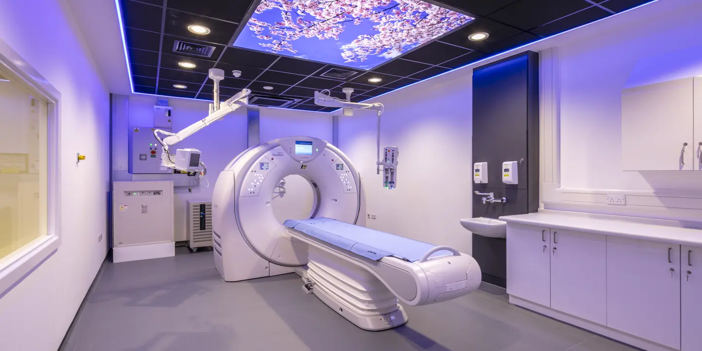
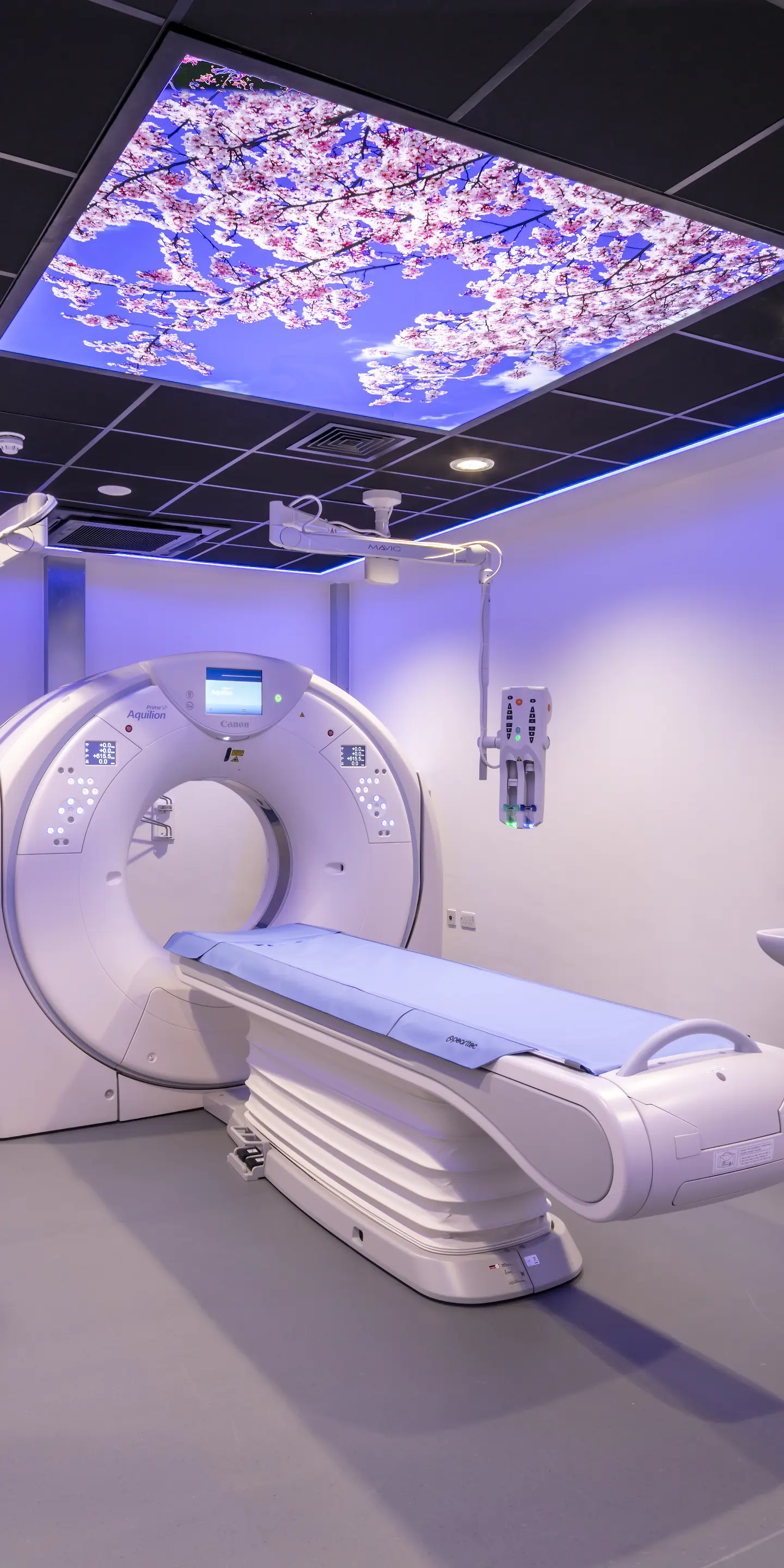
How does a CT scanner work?
During your CT thorax scan, you will be passed into a CT scanner. The 3 main parts of a CT scanner are the patient table, a ring structure and a computer. You will be asked to lie down on the patient table and then the narrow table will be slid into the ring as your scan begins.
The ring contains an X-ray generator and an X-ray detector. It gently moves around you, taking quick X-ray pictures from different angles. As the X-rays pass through your body, some are absorbed by your tissues.
Any X-rays that are not absorbed pass through your body and are picked up by the X-ray detector. This information is sent to a computer that processes it into thin image slices of your chest (cross sectional images).
As the gantry of the CT scanner moves along your body, further cross-sections are created. These cross-sections are then pieced together and converted into a 3D image.
Understanding CT images
More dense tissues, such as bone, absorb more X-rays and appear white on a CT scan image. Air absorbs very low amounts of X-rays and appears black on a CT scan image. Healthy, air-filled lungs will appear mainly black.
Other soft tissues, such as organs, muscles, ligaments and tendons, appear as varying shades of grey on CT images. In the lungs, scar tissue, fluid or growths will appear grey or white depending on their density.
Blood appears white despite not being as dense as bone. This is because it absorbs more X-rays.
Key benefits of CT thorax scans
Detailed images
As a CT thorax scan provides higher resolution images than a chest X ray, it is ideal for detecting lung, heart and other chest conditions at an early stage. Early diagnosis ensures you can get the best treatment sooner.
3D
A CT scan generates 3D images, which provide a more complete picture of the health of the organs and tissues in your chest. This helps with the accurate diagnosis of diseases and injuries.
Low radiation
Although a CT scan uses ionising radiation, the doses are low and considered to be within safe limits.
A chest CT scan uses 6.1 mSv of radiation. This is roughly equivalent to the amount of radiation you would be exposed to from the environment over 2 years.
A chest CT for lung cancer screening uses 1.5 mSv of radiation. This is about the same as what you would be exposed to from the environment over 6 months.
Speed
A chest CT scan usually takes around 15 minutes. Although this is longer than a chest X-ray, it is shorter than a chest MRI scan and produces detailed 3D images.
Conditions detected by chest CT scans
A CT thorax scan is used to investigate chest symptoms, such as difficulty breathing and chest pain. It can detect a wide range of conditions, including those affecting the lung tissues, such as:
-
Interstitial lung disease – a group of lung diseases that cause scarring of your lung tissue, specifically the tissue surrounding the air sacs (alveoli) of your lungs
-
Lung cancer
-
Pneumonia – inflammation of the lungs due to infection with bacteria, fungi or viruses
-
Pulmonary embolism – a blood clot that blocks a blood vessel supplying oxygen-rich blood to your lungs
-
Pulmonary fibrosis – thickening, stiffening and scarring of the tissue that surrounds the alveoli of the lungs
-
Tuberculosis – infection of the lungs with the bacteria Mycobacterium tuberculosis
In addition to lung conditions, a chest CT can also detect heart conditions, such as:
-
Cardiac hypertrophy – when the heart muscle becomes thicker than normal, particularly the lower left chamber
-
Heart disease
-
Heart valve defects
-
Problems with blood vessels that supply the heart
Comparing CT scans to other imaging tests
CT vs X-ray
Both a chest CT scan and a chest X-ray use ionising radiation. However, a chest CT uses about 60 times more X-rays – a chest CT scan uses 6.1 mSv of radiation while a chest X ray uses 0.1 mSv. In both cases, the amount of radiation exposure is considered to be low.
A chest X-ray is faster than a CT thorax scan, taking just a few minutes. However, it produces lower resolution images in 2D. A CT thorax scan produces more detailed images in 3D, which makes it easier to detect changes.
A chest X-ray is, therefore, often used in emergency situations as a first-line imaging tool. A chest CT is preferred when more detailed information is needed to make a definitive diagnosis
CT vs MRI
Similar to CT, MRI or magnetic resonance imaging produces detailed 3D images of the inside of your body. However, MRI does not expose you to any radiation as strong magnets and radio waves are used rather than X-rays. This makes MRI suitable for pregnant women.
Both a chest CT scan and a chest MRI scan can detect the early signs of disease. CT scans offer superior spatial resolution, which is better at highlighting the boundaries between tissues, while MRI scans offer superior contrast resolution, which is better for highlighting changes within a tissue.
An MRI scan takes longer to perform and involves being in a more enclosed space (the tube of the MRI machine). A chest MRI takes around 40 minutes – roughly double that of a CT thorax scan.
Preparing for your CT thorax scan
On the day of your scan, wear loose fitting clothing as this will make it easier for you to change into a hospital gown. You will need to remove all metal objects from your body as metal distorts the images produced. This includes metal jewellery, piercings, and hearing aids.
If your care team has said that you will need a chest CT scan with contrast, they will tell you how long you need to fast for before your scan. Contrast is a special dye that is given through a small injection into your arm. Contrast improves the clarity of the images created.
This iodine-based dye absorbs X-rays, which means any tissue with more iodine becomes brighter on a CT image. However, if you have poor kidney function, contrast may not be suitable as it can strain your kidneys.
Your care team will also advise you on whether you need to temporarily stop taking certain medications.
After your CT scan
If you have a contrast agent, you will need to remain at the scanning facility for around 30 minutes after your CT scan. You will be monitored in case you develop an allergic reaction to the contrast agent. Reactions are very rare, and most are mild if they do occur. For example, you may feel warm, and experience nausea and/or a metallic taste in your mouth.
If you do not have contrast, you can return to your usual activities immediately after your scan.
Interpreting your CT scan results and next steps
At Vista Health, we aim to deliver a detailed report of your scan results within 3 days. This report will include insights from an experienced Vista Health Consultant Radiologist.
The report can be sent to your NHS GP, or you can see one of our expert Vista Health GPs to discuss what your results mean for you, and any next steps if treatment is needed. Our team will explain your results clearly and answer any questions you may have about next steps.
Get clarity on your health today
If you are struggling with persistent chest symptoms, such as trouble breathing or chest pain, get clarity on your symptoms today with Vista Health. You can book a consultation with one of our experienced GPs who can advise you on whether a CT scan is the right scan for you.
Sources
https://www.dgft.nhs.uk/wp-content/uploads/2014/04/CT-thorax-and-upper-abdomen-V11.pdf
https://www.radiologyinfo.org/en/info/chestct
https://www.radiologyinfo.org/en/info/safety-xray
https://mft.nhs.uk/lunghealthcheck/leaflets-information/what-is-a-ct-scan/
https://www.radiologyinfo.org/en/info/chestct
https://radiologyassistant.nl/chest/pearls/cardiovascular-pearls-on-chest-ct-2
https://www.nhs.uk/tests-and-treatments/x-ray/
https://www.mdanderson.org/cancerwise/ct-scan-vs-mri--what-is-the-difference.h00-159616278.html
https://www.mdanderson.org/cancerwise/ct-scan-vs-mri--what-is-the-difference.h00-159616278.html