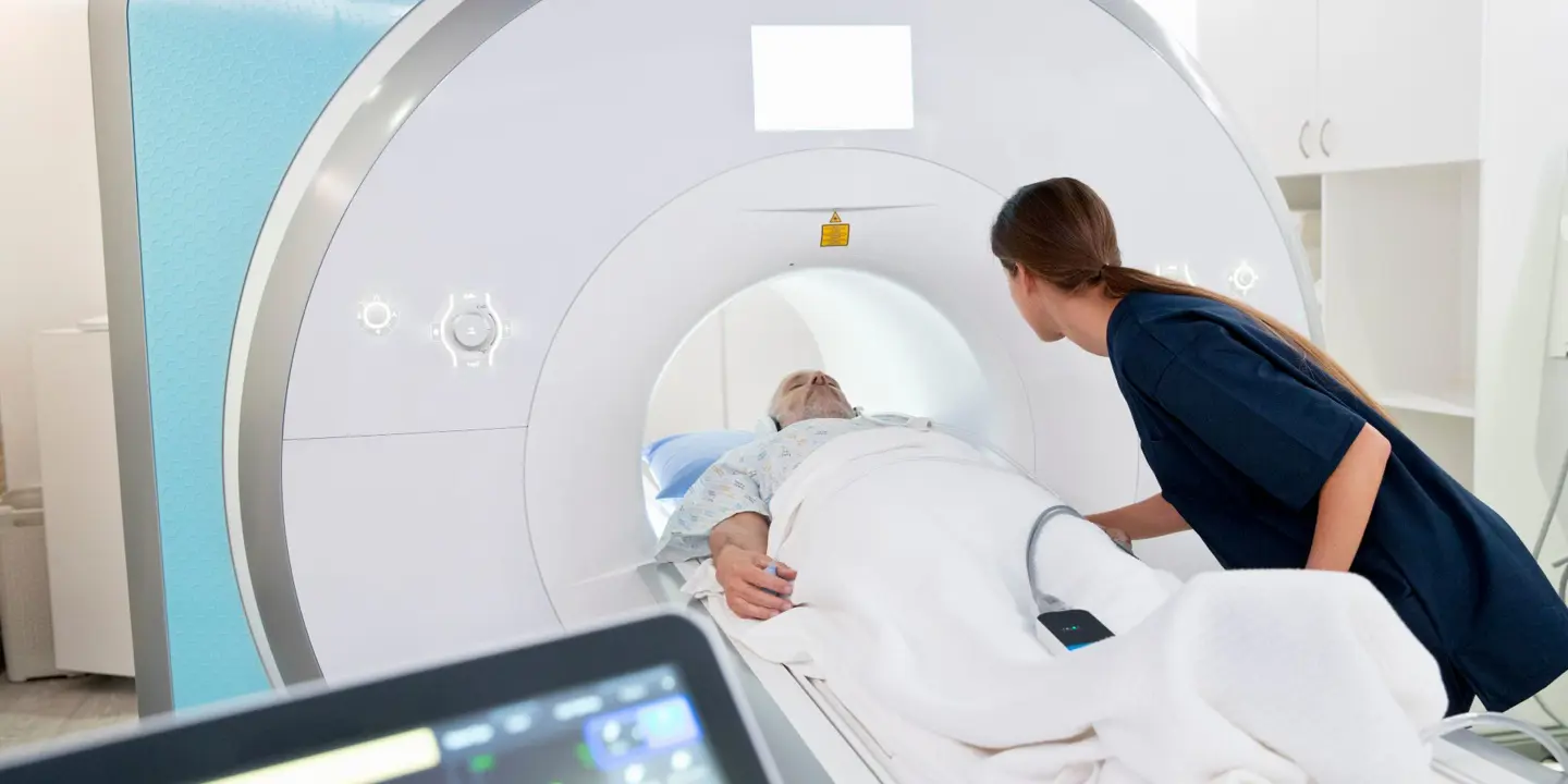
Every year, millions of people in London and across the UK have an ultrasound scan. In 2018, the NHS revealed that almost 43 million imaging tests were ordered in the U.K in a single year alone. These imaging tests included Ultrasound, as well as CT scans, and MRIs.
Ultrasound scans today, provide clinicians with images that can diagnose all sorts of conditions. But how does an ultrasound scan work? And had you realised of how it all began?
The History of Ultrasound
Using soundwaves to determine the position of objects first appeared in the 1794 work by Lazaro Spallanzani, an Italian physicist. He studied how bats managed to fly in total darkness and theorized that the nocturnal creatures were dependent on using sound to navigate.
It wasn’t until 1880, almost ninety years after Spallanzani’s theory was published that the first machine that could generate soundwaves was made. Renowned French scientists, Jacques and Pierre Curie determined that by applying electrical currents to quartz crystal they produce sound, more specifically, ultrasonic waves.
SY Sokolov, a Russian physicist, was the first to conceptualize using ultrasound for imaging techniques in 1928. However, he was more interested in using the method to find imperfections in metallic structures than for saving lives through diagnostics. However, the device he invented did create shadow images that could be interpreted.
An American scientist, George Ludwig was one of the first to use ultrasound imaging techniques. In his research about gall stones, he turned to ultrasound to detect the masses when they were embedded in soft tissue. His analysis in using these soundwaves on animal tissues helped the next scientist down the line.

It was Ian Donald who first fused ultrasound with diagnostic medicine in 1956. Naturally, his first use of the device was to determine the diameter of the head of a fetus. In only two years, technology helped create a way to visualize the findings of an ultrasound, increasing sits life-saving diagnostic potential.
We have certainly come on since George Ludvig used the apparatus shown here.
How Ultrasound Works
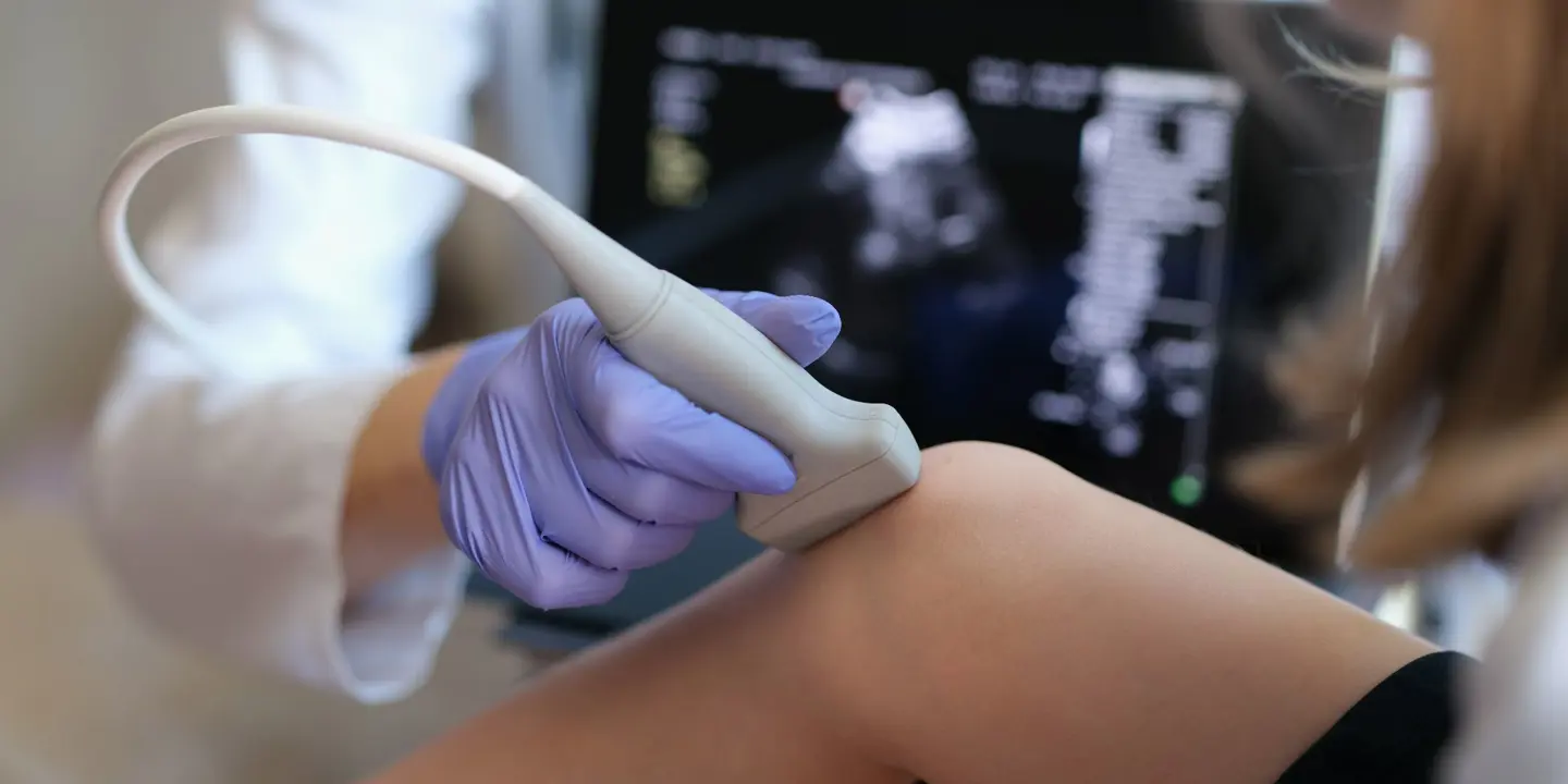
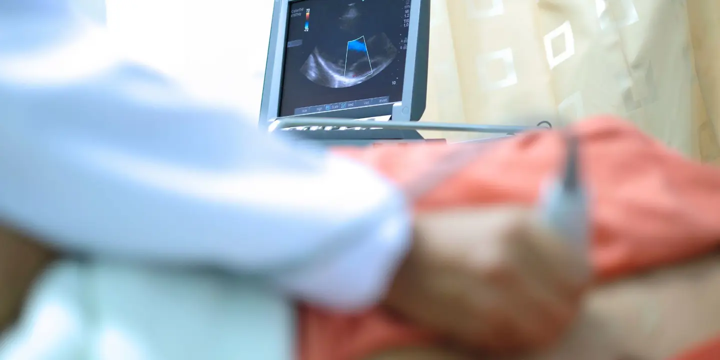
So how does an ultrasound work? The device held by an ultrasound specialist close to your body is known as a transducer. The transducer contains special crystals that can create soundwaves when they are activated with an electrical field. Not only that, but they can also receive soundwaves reflected by your body’s tissue. These tissues have different densities, which reflect varying soundwaves.
The reflected soundwaves are calculated by a computer and interpreted by a sonogram. By measuring how quickly a soundwave is reflected to the transducer, the machine can generate an image based on the data. This is shown on the screen attached to the sensor.
But what about the gel they slather on your body? What purpose does that serve? The gel makes sure there are no air pockets between your skin and the transducer. Such air pockets could block the soundwaves and prevent the ultrasound from imaging correctly.
Reliable and Affordable Medical Services
Vista Health is one of the leading providers of specialised healthcare and diagnostic services in the United Kingdom. We work with all major insurance providers and clinicians to offer our clients with quick turnarounds on assessment. We offer Private Ultrasound scans from just £125 at clinics in London and in the North West.
Enquire today on our website or call us on the number at the top of the page.