
Clinically reviewed by Hollie Phillips
National Clinical Lead & Clinical Operations Manager (Echo)
How Accurate is an Echocardiogram?
An echocardiogram is a medical imaging test that uses ultrasound to create images of the heart and its blood vessels. Also known as an echo, it is often used to help diagnose heart conditions. But how accurate is an echocardiogram?
In general, an echo is a highly accurate tool for investigating heart problems. Here, we will explore its benefits and limitations, as well as how it compares to other heart tests.
Understanding echocardiogram accuracy
Echocardiograms create detailed 2D images of your heart in real-time, which can provide highly accurate insights into your heart health. They work using ultrasound technology.
Ultrasound refers to high-frequency sound waves. These sound waves are beyond the range of human hearing. During an echo, an ultrasound probe is used to pass ultrasound waves into the body.
The sound waves are reflected as they encounter boundaries between different tissues. These reflected sound waves – called echoes – are detected by the ultrasound probe. In a single second, over 1,000 echoes can be detected. This is what enables an echocardiogram to create live images of the heart.
The echoes are converted into electrical signals and sent to a computer, where they are processed into grayscale digital images. These images reveal the structures of the heart, which includes its:
-
Four heart chambers – left ventricle, right ventricle, left atrium and right atrium
-
Four heart valves – aortic valve, mitral valve, pulmonary valve and tricuspid valve
An echo also helps assess how well the heart is functioning by imaging blood flow through the heart valves and out of the heart. This includes the evaluation of left ventricular systolic function through measurements such as the left ventricular ejection fraction (LVEF), an important indicator of how much blood the left ventricle pumps out with each contraction.
By providing information about both the structure and function of the heart at the same time, an echocardiogram is able to accurately diagnose a range of heart conditions. An abnormal echocardiogram can be a sign of the following conditions:
-
Cardiomyopathy – thickening and stiffening of the heart wall
-
Congestive heart failure – when the heart is unable to pump enough blood around the body
-
Endocarditis – inflammation of the inner lining of the heart
-
Heart disease, which is also known as coronary artery disease
-
Heart valve defects, such as aortic valve stenosis or regurgitation
-
Pericardial disease – conditions that affect the pericardium, that is, the fibrous sac-like structure that the heart sits inside
An echocardiogram can also be used to help diagnose pulmonary hypertension, that is, high blood pressure in the arteries that supply the lungs.
When to have an echo
You may want to have a private echo if you have a family history of heart problems and are, therefore, concerned about your heart health.
However, you may also choose to have a private echo if you have unexplained symptoms that may suggest a heart problem. This includes:
-
breathlessness
-
chest pain
-
feeling light-headed
-
heart palpitations ie the sensation that your heart is racing
-
persistent swelling of your ankles
-
unexplained fatigue
How is an echocardiogram performed?
The gold standard of echocardiography to investigate heart problems is a transthoracic echocardiogram (echo) or TTE. This involves passing an ultrasound probe across your chest.
When you have an echo, you will be asked to remove the clothing from your upper body.
A simple ECG, or rhythm strip is performed alongside an echo. It measures the electrical activity of your heart allowing measurements to be completed at the correct timings within your hearts cycle.
The simplified ECG is performed using 3 leads rather than a 12-lead ECG used in a standard ECG. This means 3 sticky pads (electrodes) attached to wires will be stuck to different points on your chest. The wires will connect to the Echo machine.
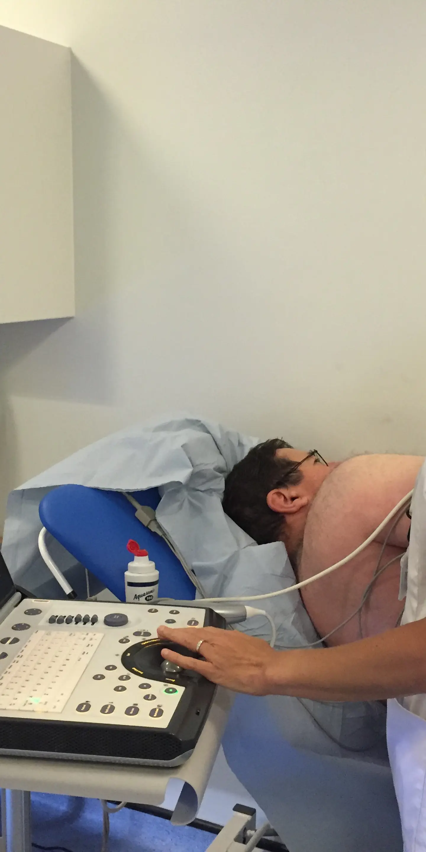
You will then be asked to lie on your left side. A gel will be applied over your chest, and the ultrasound probe will be moved across it. The test is not painful, although the gel may feel cold, and you may feel some pressure as the probe is passed across your chest.
The gel ensures transition of the ultrasound waves into your body. Any air gaps can disrupt the passage of sound waves from the probe into your body – the gel helps ensure that there are no air gaps.

Factors influencing echocardiogram results
The results of your echocardiogram can be affected by several health factors. These factors can make your echo results less clear by disrupting the passage of sound waves to the heart. They include:
-
chest deformities
-
excess fat around the heart
-
lung disease
-
obesity
If a standard echocardiogram does not provide clear enough results, your doctor may recommend a contrast-enhanced echo to enhance the precision of your echo.
Comparing echocardiograms to other tests
Echocardiogram vs ECG
An ECG measures the electrical activity of your heart. It, therefore, does not provide any visual information on the health of your heart but instead produces a graph of your heart rhythm.
In contrast, an echo provides real-time visual information of the anatomy and function of your heart, including accurate evaluation of left ventricular systolic function and parameters such as ejection fraction.
As an echo cannot provide information on your heart’s electrical activity, an ECG is better equipped to diagnose abnormalities in your heart’s electrical activity. This includes arrhythmias and heart block.
An echo, on the other hand, is better equipped to diagnose heart failure and heart valve problems.
Both an echo and an ECG can be used to detect signs that a heart attack has occurred. An echo can be performed later to assess damage to the heart.
Echocardiogram vs cardiac CT scan
A cardiac CT scan creates highly detailed images of the heart – these images are of a higher resolution than an echo. The results of the scan show the health of the heart at a single moment in time, whereas an echo provides images of the heart in real-time.
While an echo is the preferred medical imaging test for heart valve problems and heart failure, a cardiac CT scan is better at detecting problems with the blood vessels supplying the heart, namely, the coronary arteries.
A cardiac CT scan provides direct evidence of coronary artery blockages by creating high-resolution images of these blood vessels. Meanwhile, an echo provides indirect evidence of coronary artery disease, namely, the consequences of blocked coronary arteries on blood flow.
A cardiac CT scan uses ionising radiation in the form of X-rays, while an echo does not expose you to any ionising radiation.
Echocardiogram vs cardiac MRI scan
A cardiac MRI scan provides the highest resolution images of the heart. This allows cardiac MRI to detect the earliest signs of heart failure that may be missed on an echo.
However, a cardiac MRI scan is more expensive and time-consuming. It is, therefore, usually used to investigate the cause of heart failure when this is not apparent from an initial echocardiogram.
Neither an echo nor a cardiac MRI scan use ionising radiation. However, a cardiac MRI scan does use powerful magnets. This means that cardiac MRI is not suitable if you have non-MRI-compatible metal implants.
Dispelling common misconceptions about echocardiograms
An echocardiogram is not the same as an ECG
An echocardiogram is different to an ECG as it creates images of the heart in real-time while an ECG measures the electrical activity of the heart in real-time.
An echo is usually performed alongside a simple 3-lead ECG. However, depending on the results of your echo, you may still need a 12-lead ECG to gather more detailed information on the electrical activity of your heart.
An echocardiogram does not use radiation
Unlike a cardiac CT scan, which uses ionising radiation in the form of X-rays, an echocardiogram does not expose you to any ionising radiation. This is why it is completely safe to use in babies (foetal echocardiography) and pregnant women.
An echocardiogram is not painful
An echocardiogram is a safe, non-invasive and painless procedure. During a transthoracic echocardiogram, an ultrasound probe will be moved across your chest – you may feel some pressure but it is not usually painful.
Seek an expert opinion for clarity on your heart health
If you are concerned about your heart health or symptoms of a potential heart issue, you can self-refer for a private echocardiogram with Vista Health at one of our nationwide clinics.
After your scan, you will receive a full report from one of our specialist Cardiac Physiologists or Consultant Cardiologists. You will then have all the information you need to make an informed decision about your heart health.
We aim to deliver all reports within 3 working days so you can benefit from peace of mind as soon as possible.
To get clarity on your heart health, book your private echocardiogram today.
Sources
https://www.asthmaandlung.org.uk/conditions/pulmonary-hypertension/treatment
https://my.clevelandclinic.org/health/diagnostics/16947-echocardiogram
https://www.webmd.com/heart-disease/diagnosing-echocardiogram
https://www.ucsfhealth.org/medical-tests/echocardiogram
https://www.nth.nhs.uk/resources/contrast-echocardiogram-echo/
https://www.nhs.uk/conditions/heart-attack/diagnosis/
https://www.bhf.org.uk/informationsupport/tests/echocardiogram